
Image
|
Figure Caption
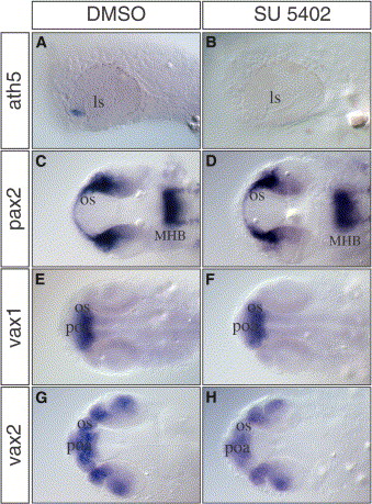
Fig. 5 Blocking Fgf Signaling Prevents the Initiation of Retina Differentiation
(A–H) (A and B) Lateral and (C–H) dorsal views of zebrafish embryos treated for 5 hours (23–28 hpf) with the (B, D, F, and H) Fgf signaling inhibitor SU5402 or (A, C, E, and G) mock solution. (A and B) In contrast to the control embryos, ath5 expression is absent in SU5402-treated embryos. Other genes expressed in the optic stalk (pax2, vax1, and vax2) were not affected by SU5402 treatment (compare [D], [F], and [H] to [C], [E], and [G], respectively). MHB, midbrain-hindbrain boundary; poa, pre-optic area.
Figure Data
Acknowledgments
This image is the copyrighted work of the attributed author or publisher, and
ZFIN has permission only to display this image to its users.
Additional permissions should be obtained from the applicable author or publisher of the image.
Reprinted from Developmental Cell, 8(4), Martinez-Morales, J.R., Del Bene, F., Nica, G., Hammerschmidt, M., Bovolenta, P., and Wittbrodt, J., Differentiation of the vertebrate retina is coordinated by an FGF signaling center, 565-574, Copyright (2005) with permission from Elsevier. Full text @ Dev. Cell