
|
Fig. 4 Fgf8 Rescues her9 and shh Expression in the oep Retina
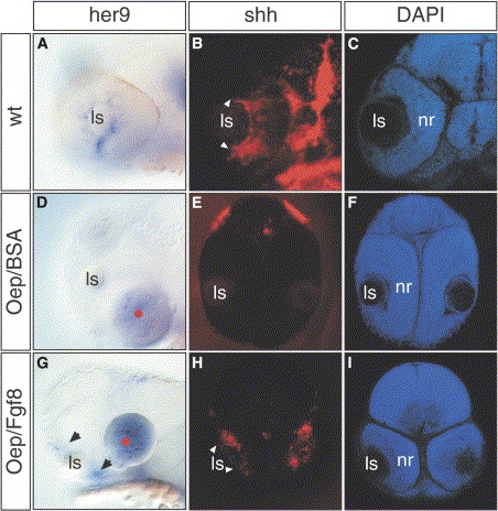
(A–I) Lateral views of (A) wild-type and (D and G) oep mutant embryos at 32 hpf. (A) her9 is normally expressed in the neuronal progenitors located in the most peripheral part of the ciliary marginal zone, close to the lens. Its expression is absent in oep mutants implanted with control beads (d, 0 out of 9), but is rescued following Fgf8 bead implantation ([G], 9 out of 10). Frontal sections of 42 hpf (B and C) wild-type and (E, F, H, and I) oep mutants after (B, E, and H) shh whole-mount in situ hybridization and (C, F, and I) DAPI counterstaining. shh is expressed in the most central part of the neural retina in a subpopulation of cells in the retinal ganglion cell and inner nuclear layers (arrowheads in [B]). This expression is absent in (E) oep control implanted embryos, but is rescued when (H) Fgf8 beads are implanted. (B and H) White arrowheads indicate the front of shh expression. (G) Black arrowheads indicate the rescue of her9 expression in the peripheral retina of oep embryos.
Reprinted from Developmental Cell, 8(4), Martinez-Morales, J.R., Del Bene, F., Nica, G., Hammerschmidt, M., Bovolenta, P., and Wittbrodt, J., Differentiation of the vertebrate retina is coordinated by an FGF signaling center, 565-574, Copyright (2005) with permission from Elsevier. Full text @ Dev. Cell