
|
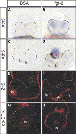
Fig. 3 Fgf8 Rescues Retinal Differentiation in oep Mutants
(A–H) (A and B) Frontal views and (C–H) coronal sections of oep embryos after (A–D) ath5 whole mount in situ staining, (E and F) Zn5, or (G and H) dp-ERK antibody staining at 28, 42, and 36 hpf, respectively. No ath5 expression is detected after implantations of BSA-soaked control beads ([A] and [C], 12 out of 12 embryos). In contrast, ath5 expression was rescued upon implantation of Fgf8-soaked beads ([B] and [D], 9 out of 11 embryos). Similarly, the RGC marker Zn5 was detected in embryos implanted with (F) Fgf8 beads, but not in those that received (E) control beads. (G and H) Dp-ERK staining was also absent in control embryos and was rescued by Fgf8 bead implantation. Dashed lines demarcate the posterior edge of the neuroretina. Rgc, retinal ganglion cells.
Reprinted from Developmental Cell, 8(4), Martinez-Morales, J.R., Del Bene, F., Nica, G., Hammerschmidt, M., Bovolenta, P., and Wittbrodt, J., Differentiation of the vertebrate retina is coordinated by an FGF signaling center, 565-574, Copyright (2005) with permission from Elsevier. Full text @ Dev. Cell