
|
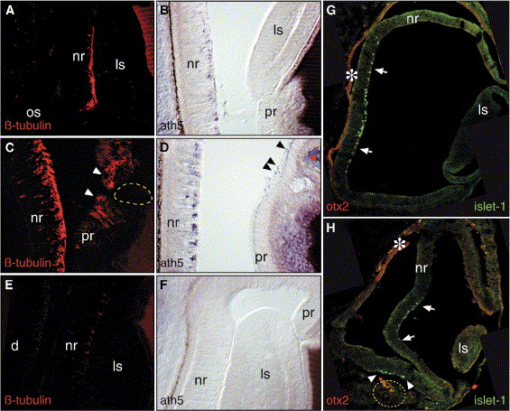
Fig. 2 Local Exposure to Fgf8 Triggers New Foci of RGC Differentiation in the Embryonic Chick Eye
(A–H) (A, B, and G) Control, (C, D, and H) FGF8, and (E and F) SU5402 bead-implanted embryos were sectioned and stained for (A, C, and E) β-tubulin, (B, D, and F) ath5, (G and H) islet-1, and Otx2 (red, labeled with an asterisk in [G] and [H]). Note that in the peripheral retina both β-tubulin and Cath5 are induced in proximity to the implanted Fgf8 bead (arrowheads in [C] and [D]). (E and F) SU5402-soaked beads inhibit neuronal differentiation. Inhibition occurs even at long range; the SU5402-soaked bead implanted is not visible within the photographed field. When beads are implanted into the periocular mesenchyme close to the RPE, Fgf8 induces an additional center of neurogenesis (arrowheads in [H]). d, diencephalon; pr, peripheral retinal.
Reprinted from Developmental Cell, 8(4), Martinez-Morales, J.R., Del Bene, F., Nica, G., Hammerschmidt, M., Bovolenta, P., and Wittbrodt, J., Differentiation of the vertebrate retina is coordinated by an FGF signaling center, 565-574, Copyright (2005) with permission from Elsevier. Full text @ Dev. Cell