
Image
|
Figure Caption
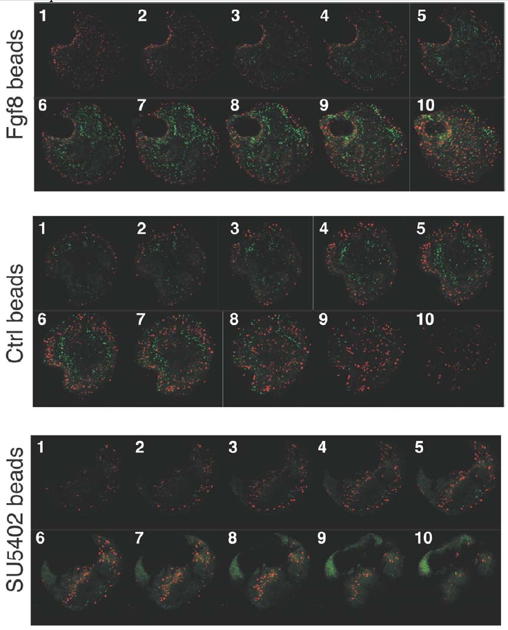
Fig. S4 A complete sequence of 10 consecutive confocal optical sections is shown for explants (Figure 1) exposed to beads soaked in Fgf8, PBS (control), and SU5402. Note the clear differences in the ratio between the numbers of differentiated cells (islet-1, green nuclei) versus proliferating cells (phospho-Histone 3, red nuclei) in those explants.
Acknowledgments
This image is the copyrighted work of the attributed author or publisher, and
ZFIN has permission only to display this image to its users.
Additional permissions should be obtained from the applicable author or publisher of the image.
Reprinted from Developmental Cell, 8(4), Martinez-Morales, J.R., Del Bene, F., Nica, G., Hammerschmidt, M., Bovolenta, P., and Wittbrodt, J., Differentiation of the vertebrate retina is coordinated by an FGF signaling center, 565-574, Copyright (2005) with permission from Elsevier. Full text @ Dev. Cell