
|
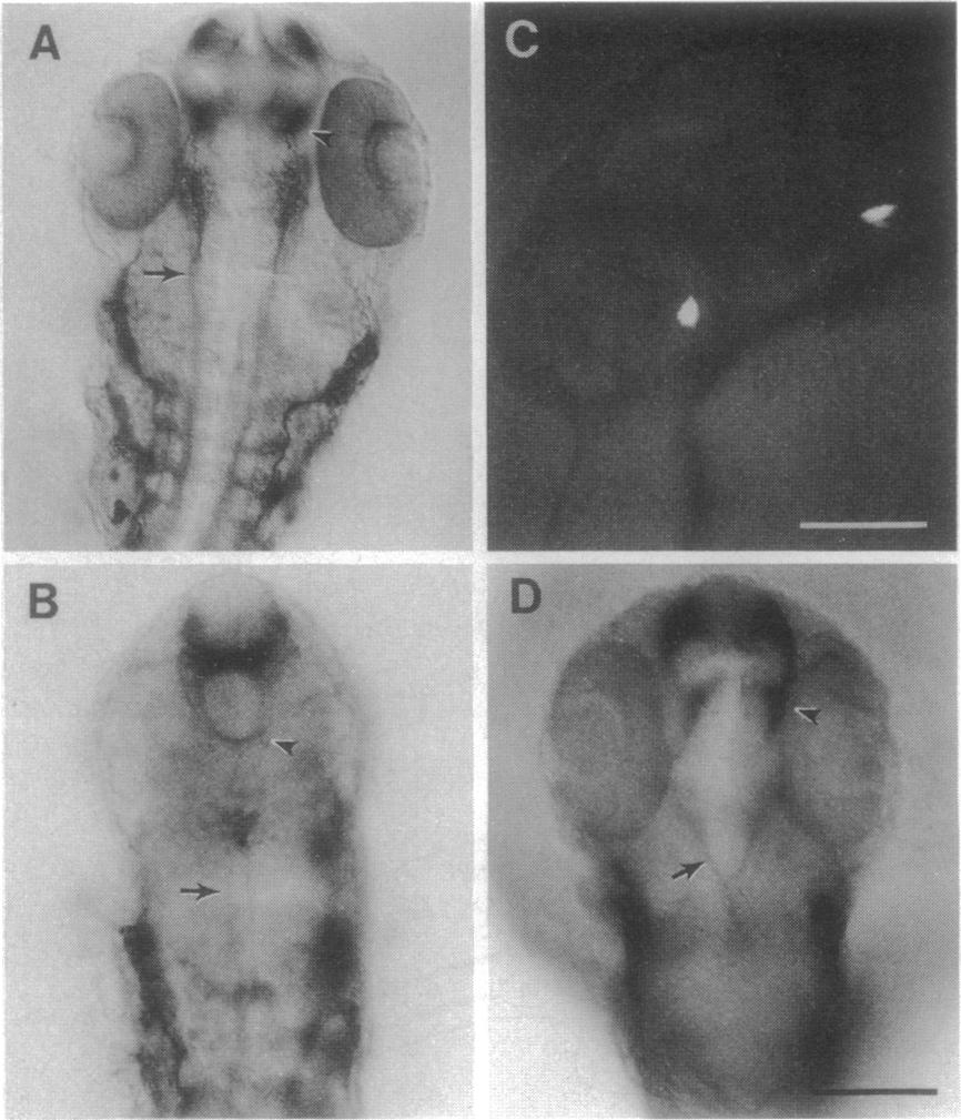
Fig. 4 Wild-type midline cells rescue diencephalic neurogenesis and axonal patterning. (A and B) Dorsal views (anterior brain at the top) of zn-12-labeled wholemounts of 26-hr wild-type (A) and cyclops (B). Normally bilaterally paired large clusters of neurons in the diencephalon (arrowhead) are completely missing in cyclops. Medial longitudinal fascicles (arrow) in the midbrain are disrupted in cyclops. (C) Left side view of the head of a living mosaic embryo, with two small clusters (ca. 2 cells each) of rhodamine-conjugated dextran- labeled transplanted wild-type cells. The cluster to the left is at the ventral midline of the diencephalon, just posterior to the eye; the other cluster is ventral to the anterior hindbrain. A few donor cells were also present in the hatching glands and lateral hindbrain but are not visible here. (D) The same mosaic embryo fixed and labeled with zn-12; the view is as in A and B. The eyes and forebrain and midbrain neuronal and axonal patterning are rescued, but not the hindbrain or spinal cord. Diencephalic neuronal clusters (arrowhead) are present; medial longitudinal fascicles (and their nuclei) are separated in the midbrain as in normal embryos but come together and fuse ventrally in the hindbrain as in mutants. (Bar = 100 μm.)