
|
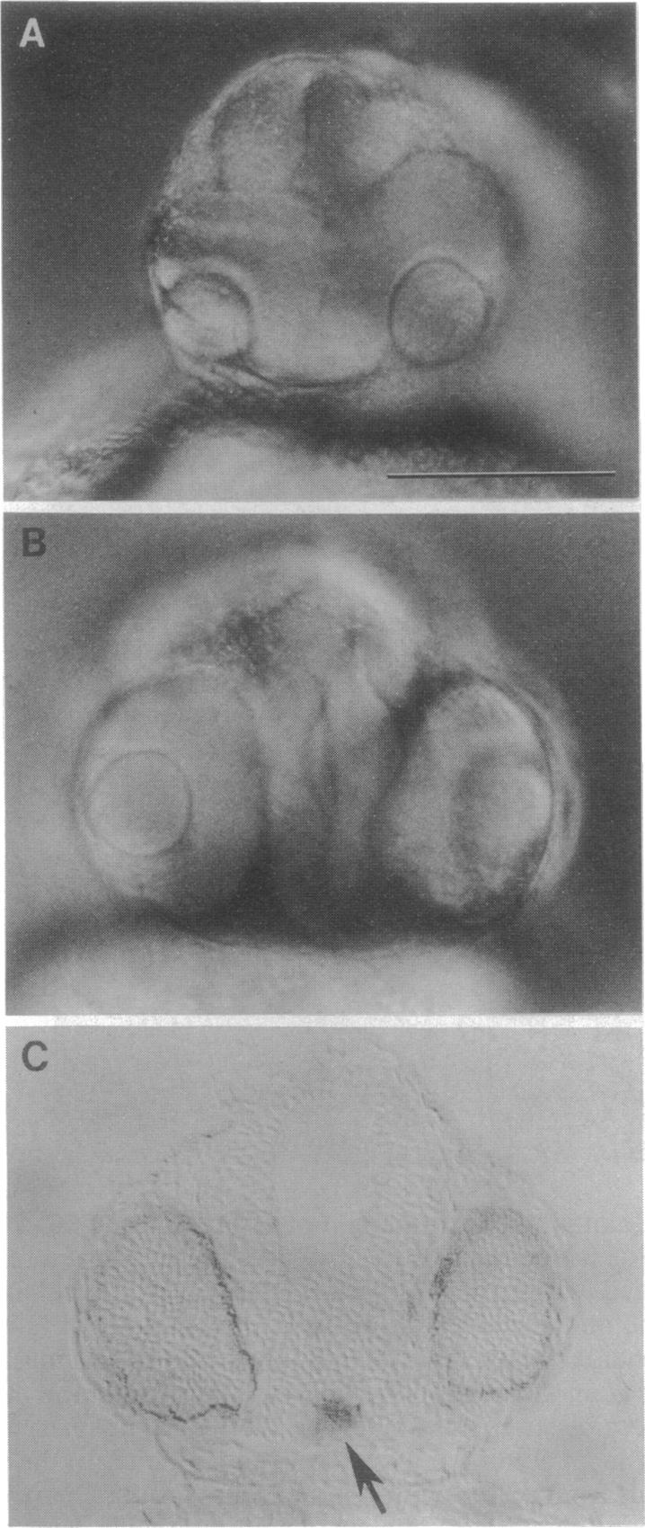
Fig. 3 Wild-type midline cells rescue cyclopia and forebrain morphogenesis in mosaics. (A) Face view of a mosaic embryo in which transplanted wild-type cells (not shown) were restricted to the trunk. The head is typically mutant in appearance. (B) Face view of a mosaic embryo in which the head morphology is essentially completely normal. However, the host was a cyclops mutant, as ascertained from the phenotype of the trunk and tail. A total of about 10 labeled wild-type cells (not shown) were present in the ventral midline of the forebrain and in the head periphery and hatching glands, which derive from the prechordal plate. (C) Transverse section through the head of another mosaic in which lateral eyes were restored (the retinal pigmented layer appears dark), and the forebrain was partially rescued by three or less biotin-labeled wild-type cells (arrow) in the midline of the ventral diencephalon. (Bar = 100 μm.)