
|
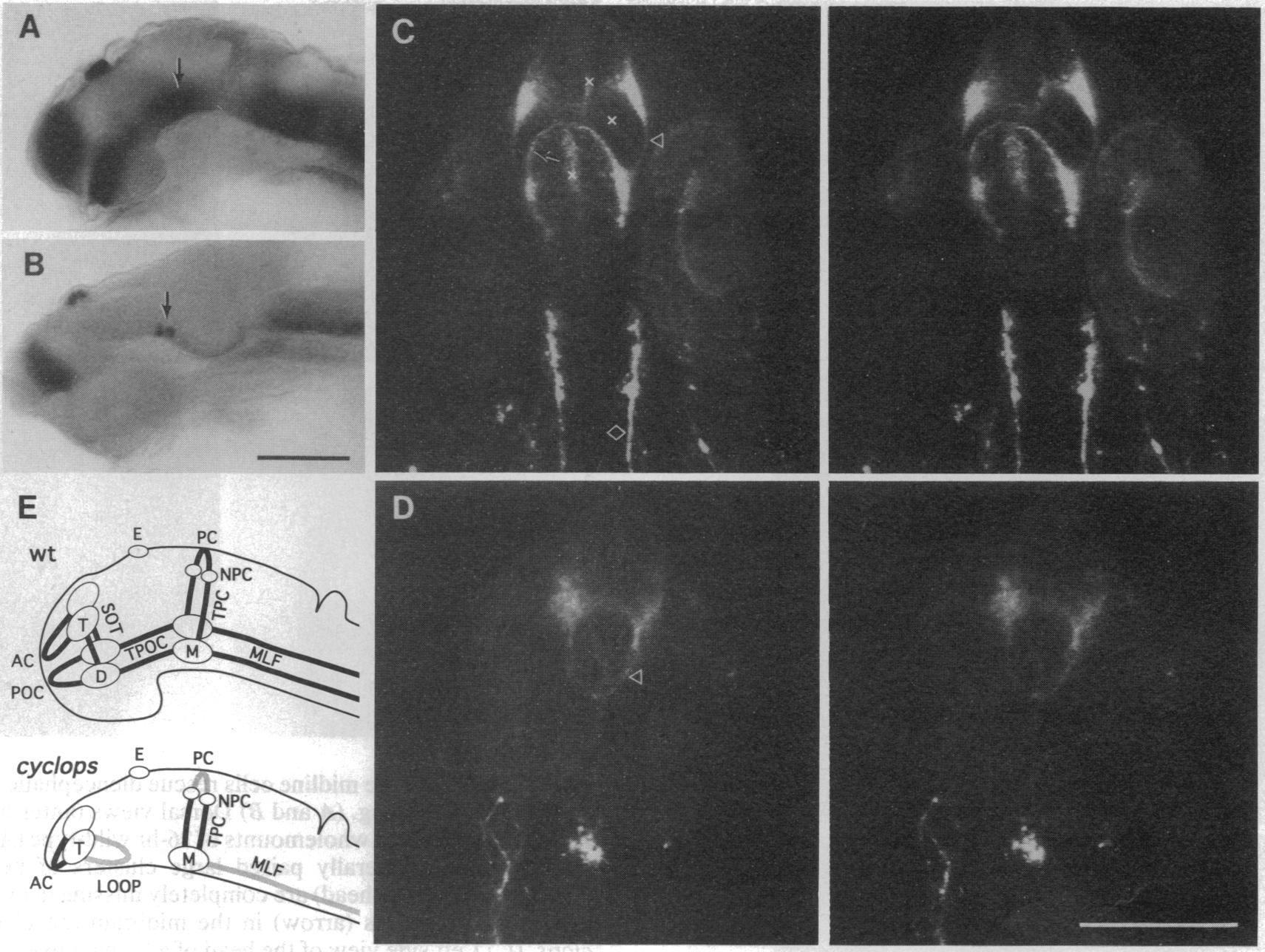
Fig. 2 cyclops alters neuronal and axonal organization in the anterior brain. Immunolabeled whole-mount preparations at 26 hr, with wild-type embryos above and mutants below. (A and B) Left side views (dorsal up) of whole-mounted wild-type embryo (A; eye removed by dissection) and mutant embryo (B; cyclopic eye present ventral to the forebrain) labeled with the monoclonal antibody zn-1, revealing neuronal cells (15). cyclops deletes the ventral diencephalic cluster and reduces the size of the midbrain cluster (arrows). The prominent telencephalic neuronal cluster (to the left) and the smaller epiphyseal cluster in the dorsal diencephalon (upper) are present in wild types and mutants alike. (C and D) Stereo-pair confocal microscopic views (from the ventral aspect, anterior brain at the top) of embryos labeled with the monoclonal antibody zn-12 (16) to show neuronal groups and axonal pathways. The postoptic commissure is prominent in the wild type (arrow in C) but is missing in the mutant (D), and the supraoptic tracts (arrowheads) connect together in an abnormal loop across the midline. In the wild type, the medial longitudinal fascicles form prominent posteriorly directed tracts (diamond) from paired midbrain neuronal groups, but these tracts are absent in this example of the mutant, and the midbrain neurons occupy a single midline cluster. x, Labeling of neuroepithelial cells, not axonal fascicles. (E) Left-side summary views of the neuronal and axonal deficiencies. Tracts that are variably present or absent in cyclops are indicated by gray lines. In cyclops, the anterior commissure (AC) is shortened, the supraoptic tracts (SOT) frequently (in 70% of the cases, n = 24) form an abnormal commissural loop posterior to the fused optic stalk. The posterior commissure (PC) was present in 40% of the mutants (n = 24). D, ventral diencephalic neuronal cluster; E, epiphyseal cluster; M, midbrain cluster; MLF, medial longitudinal fascicle; NPC, nucleus of the posterior commissure; POC, postoptic commissure; T, telencephalic cluster; TPC, tract of the posterior commissure; TPOC, tract of the postoptic commissure. (Bars = 100 μm.)