
|
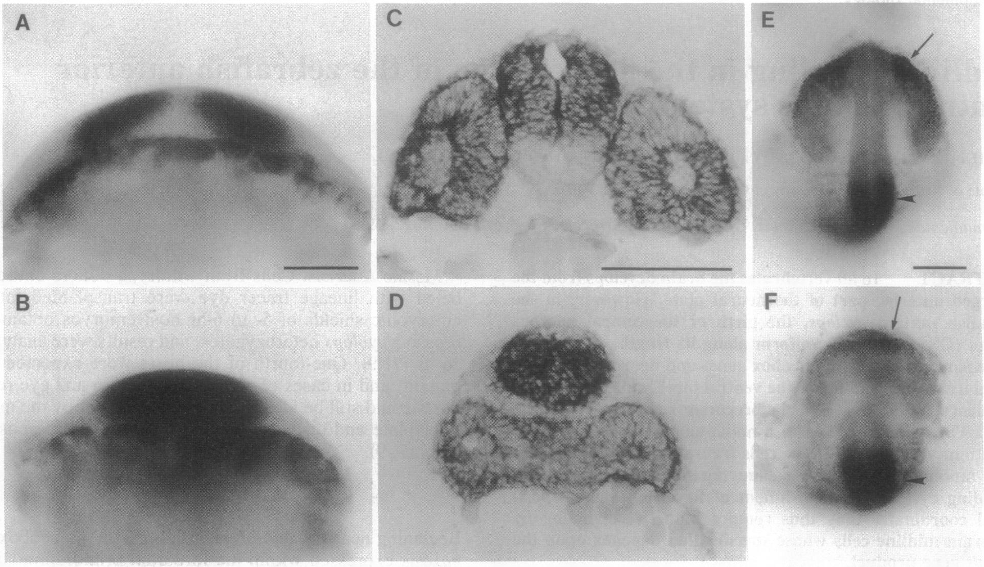
Fig. 1 cyclops alters the patterns of pax6 (A-D; in situ hybridization) and pax2 (E and F; immunolabeling) expression in the forebrain. (Upper) Wild-type embryos. (Lower) Matching homozygous cyclops mutants (cycb16; ref. 7). (A) Frontal view of whole mount at 11 hr postfertilization at 28.5°C showing the forebrain region of the wild-type neural plate. Two bilateral domains of pax6 expression are separated by a median area free of expression. (B) In cyclops, a single domain spans the midline. (C) Transverse section at 24 hr of the wild-type forebrain and eyes. pax6 expression is present in the retinas and along about two-thirds of the dorsolateral walls of the diencephalon. Expression is absent in a large ventral region and in the roof plate. (D) In cyclops, the area normally devoid of pax6 in the ventral diencephalon is entirely missing, and the retinas are fused under the diencephalon. The roof plate remains free of expression. (E) Dorsal view of the head of a wild-type whole mount at 18 hr immunolabeled with anti-Pax2 antibody. Anterior brain is at the top. Cell nuclei are labeled in bilateral patches around the anterior edges of the optic stalks (arrow) and in a band at the presumptive midbrain-hindbrain junction area (arrowhead). (F) In cyclops, the two domains of labeling in the optic stalks are weaker and are fused ventromedially (arrow). There is no obvious change in labeling at the midbrain-hindbrain junction (arrowhead). (Bar = 100 μm.)