
|
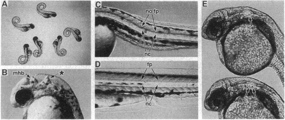
Fig. 2 Embryonic mutants recovered from backcross screens. Mutations initially identified in backcrosses between F2 offspring and their F1 parents were propagated by crossing F2 offspring to wild-type mates to generate F3 carriers. All mutant embryos depicted were generated by intercrossing F3 carriers except for the heterozygote shown in E, which was generated by crossing an F3 carrier to a wild-type mate. For embryos in B-E, anterior is to the left and dorsal is up. (A) Several 48-h embryos that are homozygous for a recessive lethal mutation causing dorsal curvature of the tail. Their shared abnormalities constitute a clearly defined syndrome. (B) A 30-h embryo that is homozygous for a recessive lethal mutation affecting brain patterning. The mutant displays a well-formed midbrain-hindbrain border (mhb) and an abnormal structure (*) in the middle of the hindbrain. (C) Trunk region of a 48-h embryo that is homozygous for a recessive lethal mutation that perturbs neuraxial development. The mutant displays a relatively normal notochord (nc) and a slightly reduced spinal cord, but the floor plate (fp) of the spinal cord is not present. (D) Trunk region of a 48-h wild-type embryo showing the notochord (nc) and the floor plate (fp) of the spinal cord. (E) A 24-h wild-type embryo (lower embryo) and a mutant sibling (upper embryo) that is heterozygous for a recessive lethal mutation with a dominant defect in inner ear development. In the wild-type embryo, two otoliths (arrows) are clearly visible in the inner ear, one in the future utricle (anterior) and another in the future saccule (posterior). In contrast, the mutant embryo lacks the anterior otolith. Heterozygotes are rarely affected in both ears and usually grow to adulthood normally. Homozygotes usually show bilateral loss of utricular otoliths, display severe equilibrium deficits, and die by day 10 of larval development. Saccular otoliths are always present in both heterozygotes and homozygotes.