
Image
|
Figure Caption
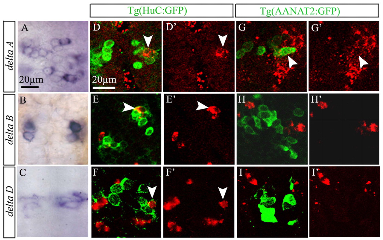
Fig. 2 deltaB and deltaD are expressed specifically in projection neurons. (A-C) Dorsal view of epiphysis, showing expression of deltaA, deltaB and deltaD in wild-type embryos at 18 hours. (D-F′) Confocal section of the epiphysis, showing expression of deltaA, deltaB and deltaD (red) in Tg(HuC:GFP) embryos at 24 hours. (G-I′) Confocal section of the epiphysis, showing expression of deltaA, deltaB and deltaD (red) in Tg(AANAT2:GFP) embryos at 24 hours. Anterior is upwards. Scale bars: 20 μm. White arrowheads indicate double-labeled cells.
Figure Data
Acknowledgments
This image is the copyrighted work of the attributed author or publisher, and
ZFIN has permission only to display this image to its users.
Additional permissions should be obtained from the applicable author or publisher of the image.
Full text @ Development