
Image
|
Figure Caption
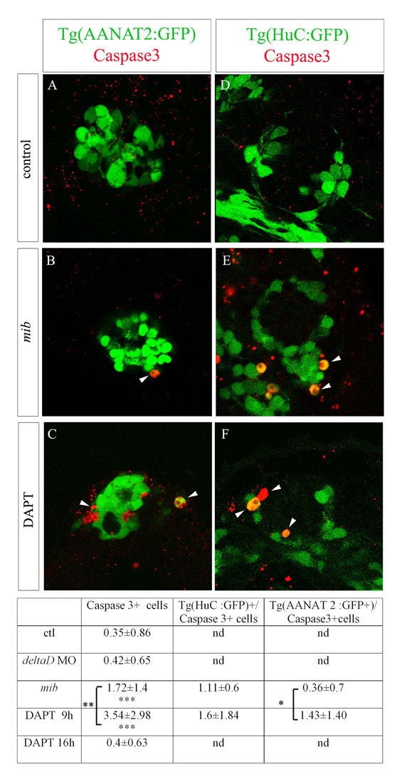
Fig. S2 (A-F) Confocal sections of Tg(AANAT2:GFP) (A-C) and Tg(HuC:GFP) (D-F) epiphysis stained with a caspase 3 antibody and GFP in wild-type (A), mib mutant (B) and DAPT-treated embryos (C) at 48 hours of development. The number of apoptotic cells is increased in mib mutant compared with wild-type embryos and in DAPT-treated embryos compared with mib mutants. (G) Counts of caspase 3+ cells, Tg(HuC:GFP)+/caspase 3+ and Tg(AANAT2:GFP)+/caspase 3+ cells. Numbers indicate average±s.d. *P<0.05; **P<0.001; ***P<0.0005 using a t-test. nd, not determined.
Acknowledgments
This image is the copyrighted work of the attributed author or publisher, and
ZFIN has permission only to display this image to its users.
Additional permissions should be obtained from the applicable author or publisher of the image.
Full text @ Development