
Image
|
Figure Caption
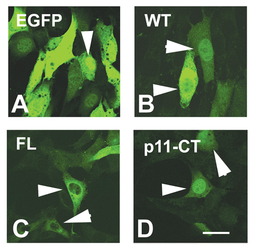
Fig. S3 Subcellular localization of Cx55.5 protein variants in transfected NIH3T3 cells: Confocal laser scanning imaging of the subcellular distribution of EGFP and the Cx55.5-EGFP fusion protein constructs in NIH3T3 cells. This experiment confirmed the distinct distribution of FL and p11-CT isoforms. Note that p11-CT showed a pronounced nuclear accumulation with some staining in the cytoplasm. Scale bar: 25 μm.
Acknowledgments
This image is the copyrighted work of the attributed author or publisher, and
ZFIN has permission only to display this image to its users.
Additional permissions should be obtained from the applicable author or publisher of the image.
Open Access
Full text @ BMC Mol. Biol.