
Image
|
Figure Caption
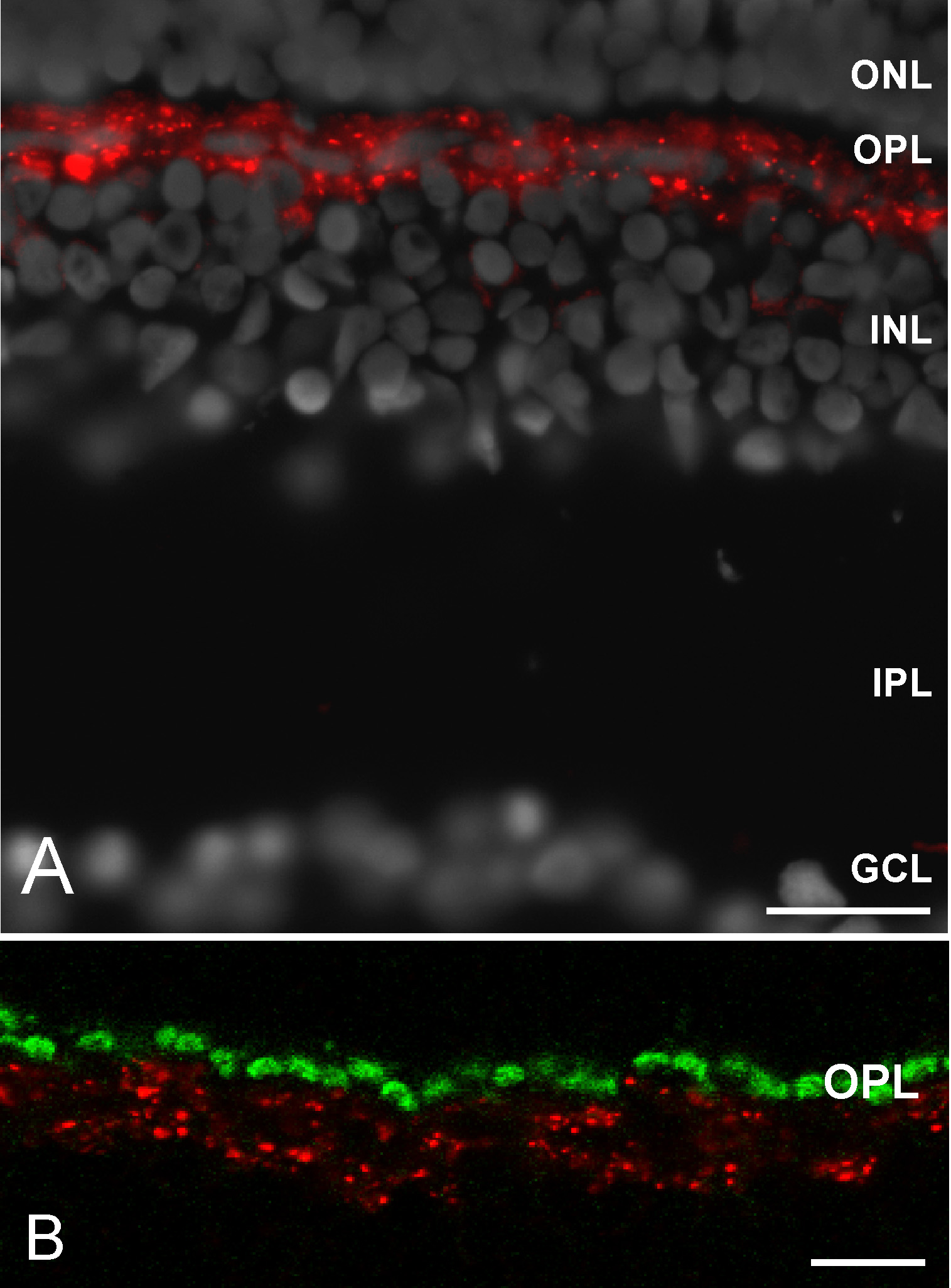
Fig. S1 Cx55.5 protein localization in the outer retina of the zebrafish. (A) Overview of the retina demonstrating the highly restricted expression of Cx55.5 in a single band like cell layer. (B) The higher magnification demonstrates the co-localization of Cx55.5 and GluR2 proteins in horizontal cells. (Abbreviations: ganglion cell layer, GCL; inner plexiform layer, IPL; inner nuclear layer, INL; outer plexiform layer, OPL; outer nuclear layer, ONL; pigment epithelium, PE). Scale bar in Fig A = 20 μm. Scale bar in B = 10 μm.
Figure Data
Acknowledgments
This image is the copyrighted work of the attributed author or publisher, and
ZFIN has permission only to display this image to its users.
Additional permissions should be obtained from the applicable author or publisher of the image.
Open Access
Full text @ BMC Mol. Biol.