
|
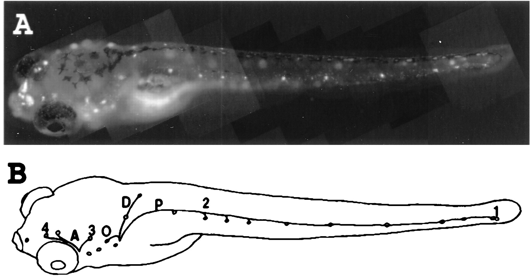
Fig. 1 (A) Vitally labeled neuromast hair cells in a 6-day-old zebrafish larvae. The dye also is taken up by the nasal epithelium. (B) Neuromasts and lateral line nerve branches whose projection was examined in the present work. Neuromasts on one side of the fish are drawn as small circles. The nerve branches that were not back-filled in this work are not shown. A, anterior lines; P, midbody posterior line; D, dorsal line; O, occipital line; 1, last two neuromasts of the posterior midbody line; 2, second neuromast of the posterior midbody line; 3, neuromast located dorso anterior to the ear vesicle; 4, last neuromast of the supra orbital line. Neuromasts 1 and 2 are innervated by the posterior lateral line ganglion, and neuromasts 3 and 4 are innervated by the anterior lateral line ganglion. Magnification: x20.