
Fig. 1
- ID
- ZDB-IMAGE-080617-13
- Publication
- Aamar et al., 2008 - Protocadherin-18a has a role in cell adhesion, behavior and migration in zebrafish development
- All Figures
- Figures for Aamar et al., 2008
|
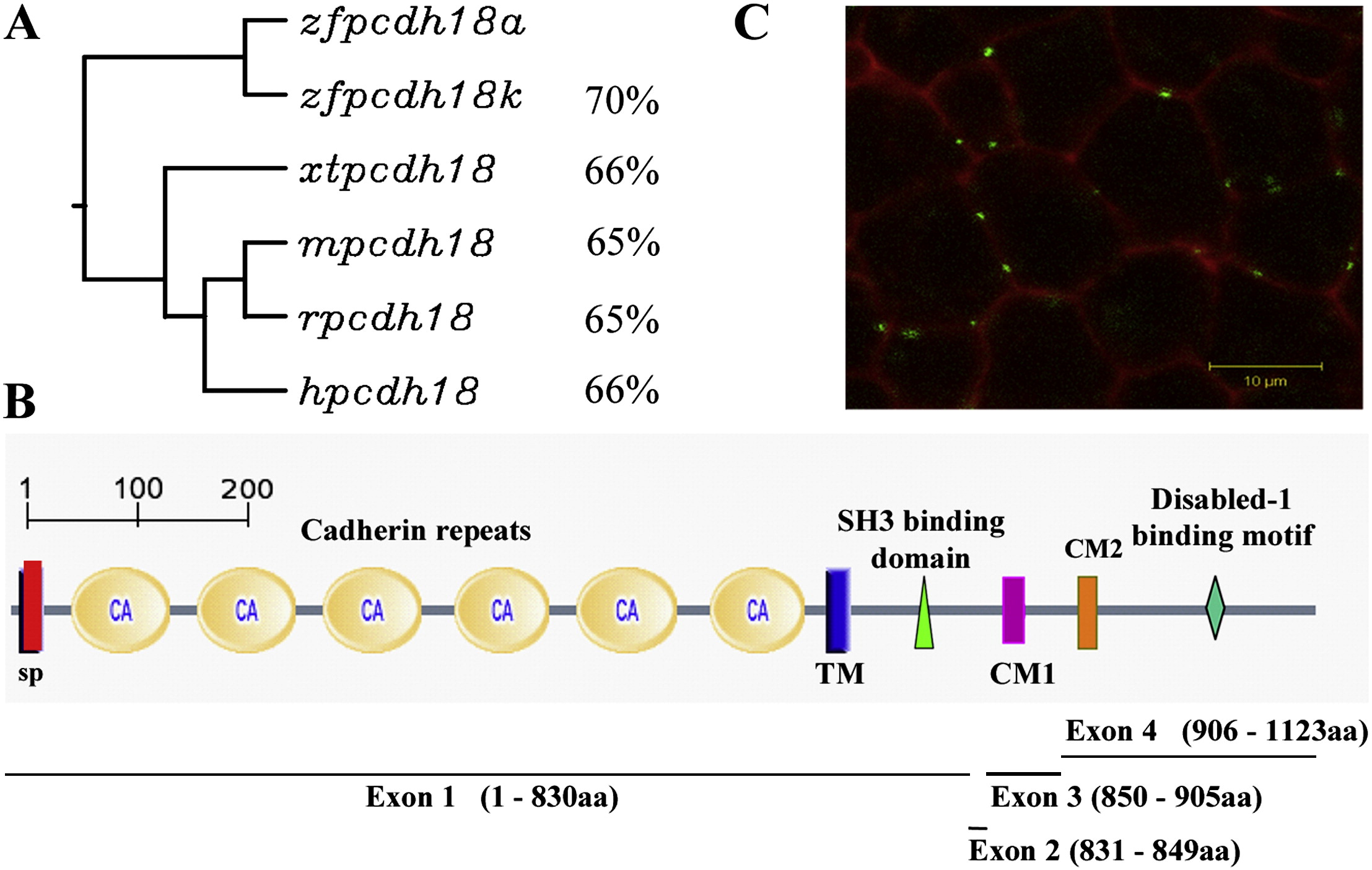
Fig. 1 Pcdh18 family homology, predicted structure and localization. (A) The closest relative to the protein we describe, Pcdh18a (ABX64360), is the protein described by (Kubota et al., 2008), labeled Pcdh18k (BAF96395). Both are related to Pcdh18 in Mus musculus (m, AAL47095, 65%), Homo sapiens (h, AAH93815, 66%), Xenopus tropicalis (xt, NP_001011150, 66%) and Rattus norvegicus (r, XP_227117, 65%). (B) Predicted protein structure of zebrafish Pcdh18a. SP, signal peptide sequence (1–29aa); CA1–CA6, six cadherin repeats in the extracellular region (30–701aa); TM, transmembrane region (702–724aa); the cytoplasmic tail (725–1123aa) contains two conserved motifs, CM1 (858–888aa) and CM2 (909–926aa), an SH3 binding motif (755–758aa) and a Disabled-1 binding motif (1044–1047aa). Below is shown the exon/intron arrangement of zfpcdh18a, containing four exons as is typical of pcdhs. (C) Confocal image of a zebrafish embryo at 80% epiboly injected at the one-cell stage with membrane-bound red fluorescent protein (mRFP, red) and pcdh18a-eGFP fusion construct (green). The fusion protein is expressed in a punctate manner at the membranes between cells. The scale is 10 μm.
Reprinted from Developmental Biology, 318(2), Aamar, E., and Dawid, I.B., Protocadherin-18a has a role in cell adhesion, behavior and migration in zebrafish development, 335-346, Copyright (2008) with permission from Elsevier. Full text @ Dev. Biol.