
|
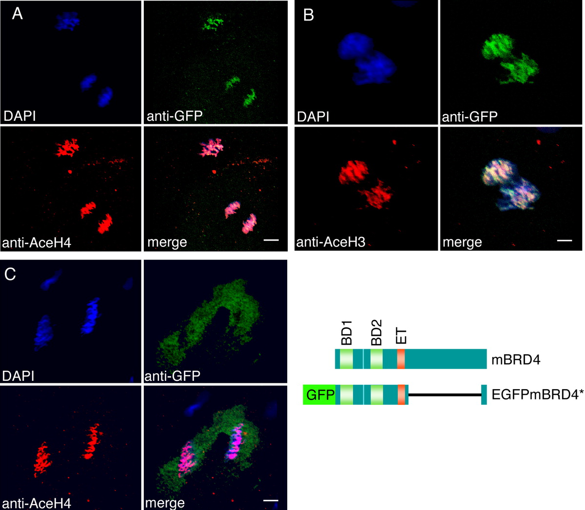
Fig. 6 Brd4 protein is colocalized with acetylated histones in zebrafish embryos. A-C: Enhanced green fluorescent protein (EGFP) -mBrd4* RNA lacking amino acids 700-1317 was injected into one- to two-cell embryos (A,B); EGFP RNA was injected as control in (C). The embryos were fixed before midblastula transition (MBT) at 256-cell (A,C) and 128- to 256-cell stage (B), and immunostained with anti-acetyl-histone antibodies (red) and anti-GFP antibodies (green). A: Anti-tetra-acetyl-histone H4; (B) anti-di-acetyl-histone H3; (C) anti-tetra-acetyl-histone H4. The structures of wild type and fusion brd4 constructs are shown. Scale bar = 8 μm in A,C; 4 μm in B.