
IMAGE
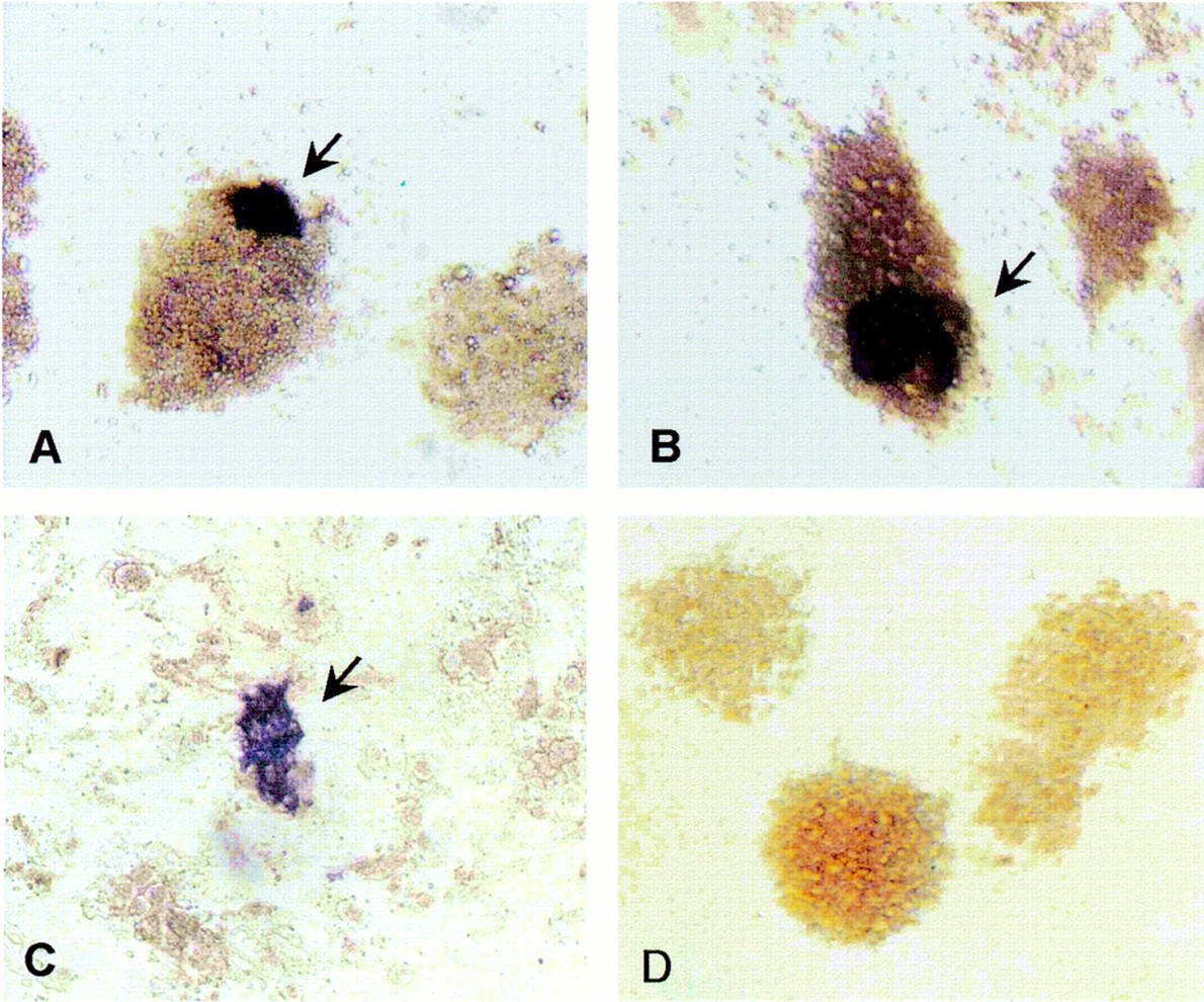
Fig. 3
- ID
- ZDB-IMAGE-080606-3
- Publication
- Ma et al., 2001 - Production of zebrafish germ-line chimeras from embryo cell cultures
- All Figures
- Figures for Ma et al., 2001
Image
|
Figure Caption
Fig. 3 Distribution of vasa-positive embryo cells. Cultures maintained for 3 days (a) and 8 days (b) in RTS34st cell-conditioned medium or 3 days on RTS34st feeder cells (c) were examined by in situ hybridization by using a vasa-specific antisense probe. The control culture (d) was grown for 8 days in conditioned medium and hybridized with sense probe. [Magnification = x200 (a, b, and d), and = x100 (c).]
Acknowledgments
This image is the copyrighted work of the attributed author or publisher, and
ZFIN has permission only to display this image to its users.
Additional permissions should be obtained from the applicable author or publisher of the image.
Full text @ Proc. Natl. Acad. Sci. USA