
Fig. S10
- ID
- ZDB-IMAGE-080605-9
- Genes
- Antibodies
- Publication
- Sidi et al., 2008 - Chk1 Suppresses a Caspase-2 Apoptotic Response to DNA Damage that Bypasses p53, Bcl-2, and Caspase-3
- All Figures
- Figures for Sidi et al., 2008
|
Fig. S10
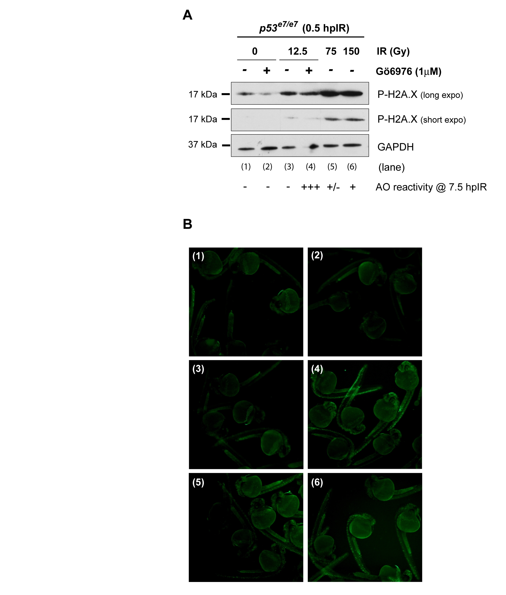
Extreme IR-induced DNA damage fails to force apoptosis in zebrafish p53 mutants endowed with wild-type Chk1 activity
(A) Western blot comparing the levels of phosphorylated H2A.X in protein lysates from p53 mutant embryos 0.5 hr after 0, 12.5, 75 or 150 Gy IR in the presence or absence of the specific Chk1 inhibitor Gö6976 (1 μM). Acridine orange (AO) reactivity at 7.5 hpIR of embryos from the same experiment is indicated below the blot (see panel B for images of representative embryos). Note that AO reactivity does not correlate with levels of DNA damage. Specifically, IR doses up to 150 Gy (which lead to dramatic levels of DNA damage) are insufficient to mimic the combinatory effects of 12.5 Gy + Chk1 inhibitor treatment.
(B) Fluorescence images of AO-stained embryos from the experiment described in A. Corresponding western blot lanes are indicated in upper left corners.
Reprinted from Cell, 133(5), Sidi, S., Sanda, T., Kennedy, R.D., Hagen, A.T., Jette, C.A., Hoffmans, R., Pascual, J., Imamura, S., Kishi, S., Amatruda, J.F., Kanki, J.P., Green, D.R., D'Andrea, A.A., and Look, A.T., Chk1 Suppresses a Caspase-2 Apoptotic Response to DNA Damage that Bypasses p53, Bcl-2, and Caspase-3, 864-877, Copyright (2008) with permission from Elsevier. Full text @ Cell