
|
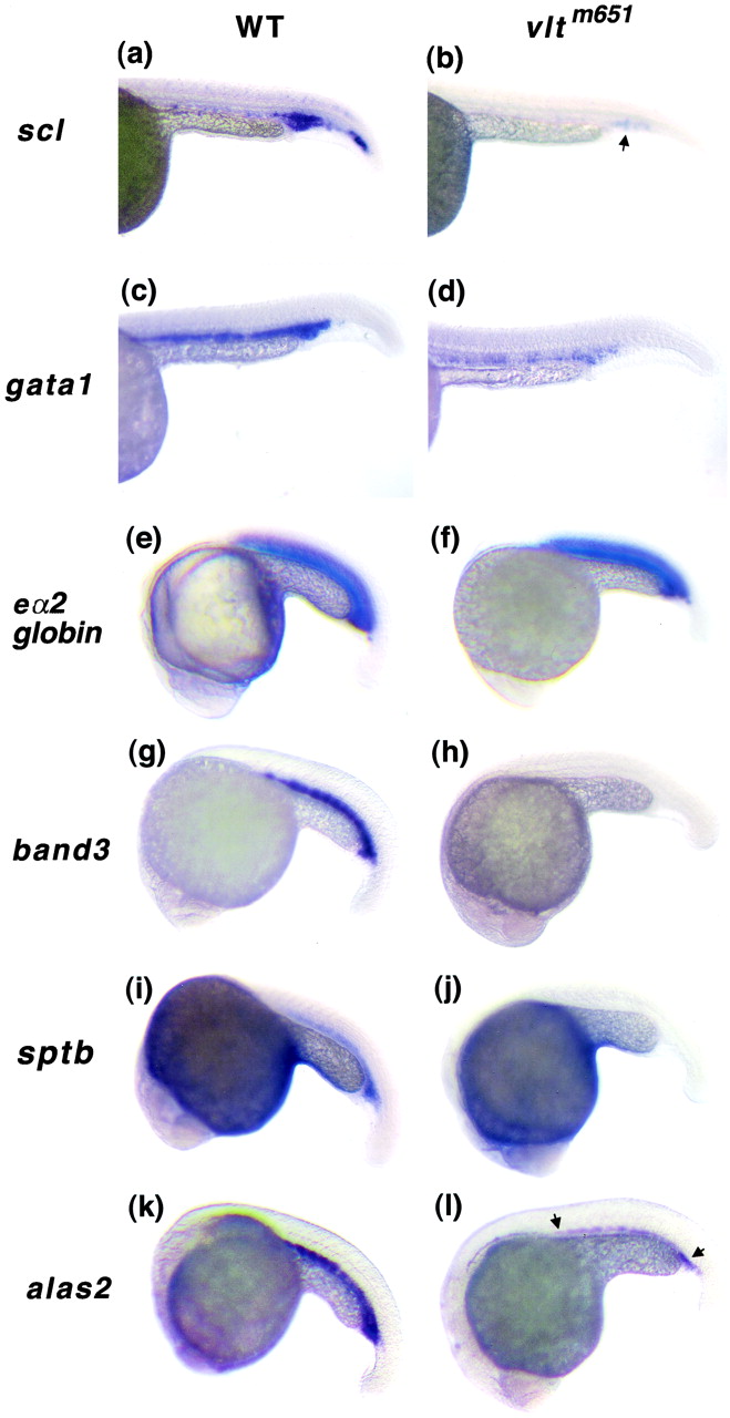
Fig. 1 Expression of hematopoietic genes in vltm651 by whole-mount RNA in situ hybridization. Embryos from vltm651 heterozygote incrosses are shown in lateral views with the heads to the left. Embryo stages are 26 hpf (a-d) and 21 somites (e-l). In situ analyses were performed on embryos from at least three independent crosses. Wild-type sibling embryos (WT) are shown in a, c, e, g, i, and k, and mutant embryos (vltm651) are shown in b, d, f, h, j, and l with scl (a and b), gata1 (c and d), globin eα2 (e and f), band3 (g and h), sptb (i and j), and alas2 (k and l) RNA probes. An arrow in b shows residual staining in the posterior ICM with scl. Arrows in l demarcate the reduced staining in the ICM with alas2.