
Fig. S1
- ID
- ZDB-IMAGE-080605-1
- Publication
- Sidi et al., 2008 - Chk1 Suppresses a Caspase-2 Apoptotic Response to DNA Damage that Bypasses p53, Bcl-2, and Caspase-3
- All Figures
- Figures for Sidi et al., 2008
|
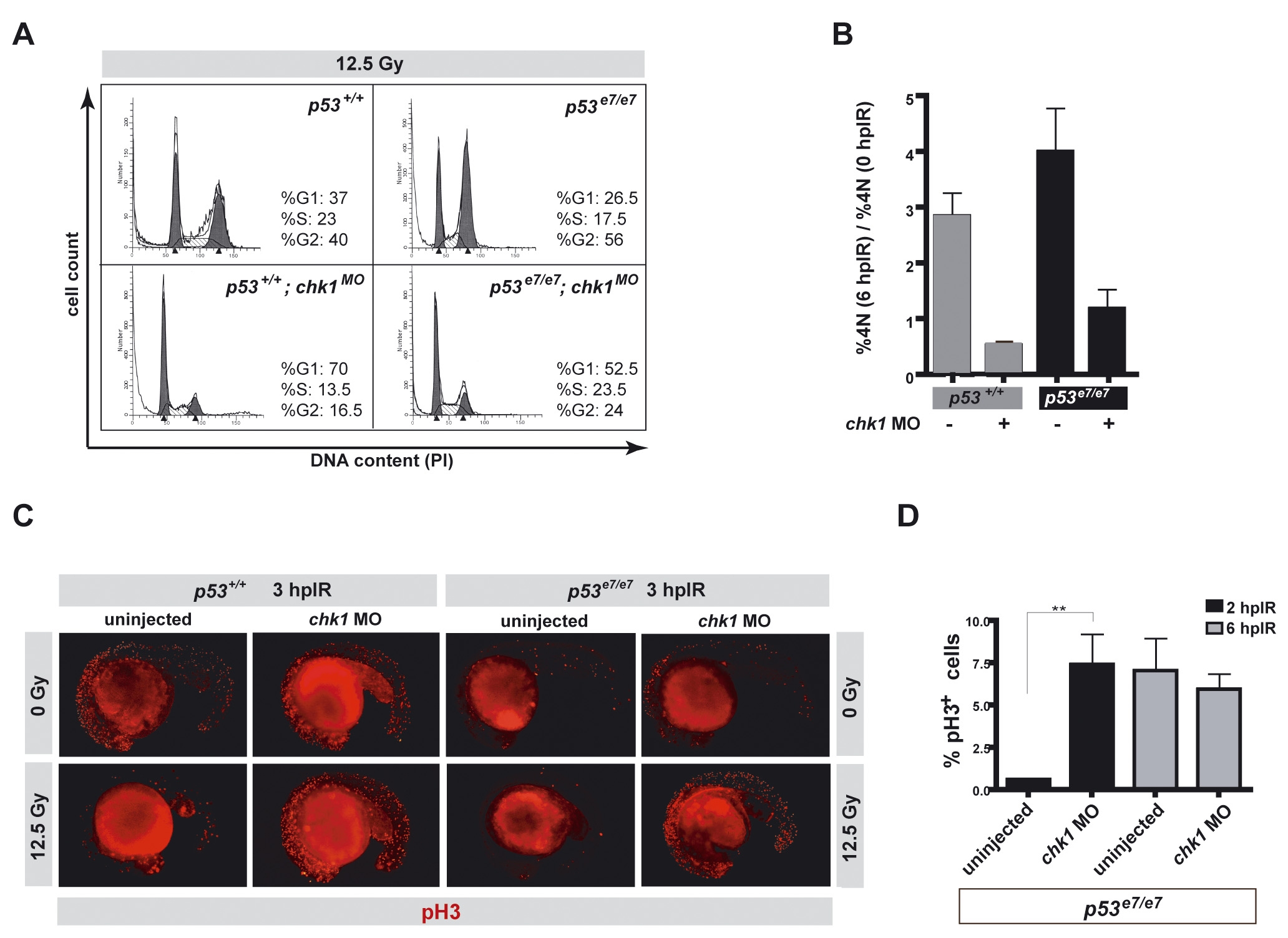
Fig. S1
. Chk1 depletion eliminates the IR-induced G2/M checkpoint in zebrafish
(A) Cell-cycle profiles of zebrafish embryos of the indicated genotypes (6 hpIR) as determined by flow cytometry. DNA content analyzed by PI staining. Note that cells from irradiated chk1 morphants fail to accumulate in G2 at 6 hpIR, regardless of p53 status.
(B) 4N DNA ratios (6 hpIR / 0 hpIR) as determined by flow cytometry of PI-stained whole embryo homogenates. Embryos were irradiated (12.5 Gy) at 18 hpf. Data collected from 3 independent experiments are reported as means ± SD.
(C) Fluorescence images of representative embryos of indicated genotypes immunostained with an anti-phospho histone H3 antibody. Note that chk1MO embryos analyzed at 2 hpIR show dramatically increased numbers of phospho-histone H3 (pH3)-positive (mitotic) cells compared with chk1WT embryos, again irrespective of their p53 genotype.
(D) Quantification of the pH3 immunostainings shown in panel C. Percentages of pH3 positive cells are means ± SEM. ** P<0.01 (two-tailed Student’s t-test). Note that the mitotic phenotype of chk1MO embryos was only transient, consistent with a checkpoint defect.
Reprinted from Cell, 133(5), Sidi, S., Sanda, T., Kennedy, R.D., Hagen, A.T., Jette, C.A., Hoffmans, R., Pascual, J., Imamura, S., Kishi, S., Amatruda, J.F., Kanki, J.P., Green, D.R., D'Andrea, A.A., and Look, A.T., Chk1 Suppresses a Caspase-2 Apoptotic Response to DNA Damage that Bypasses p53, Bcl-2, and Caspase-3, 864-877, Copyright (2008) with permission from Elsevier. Full text @ Cell