
|
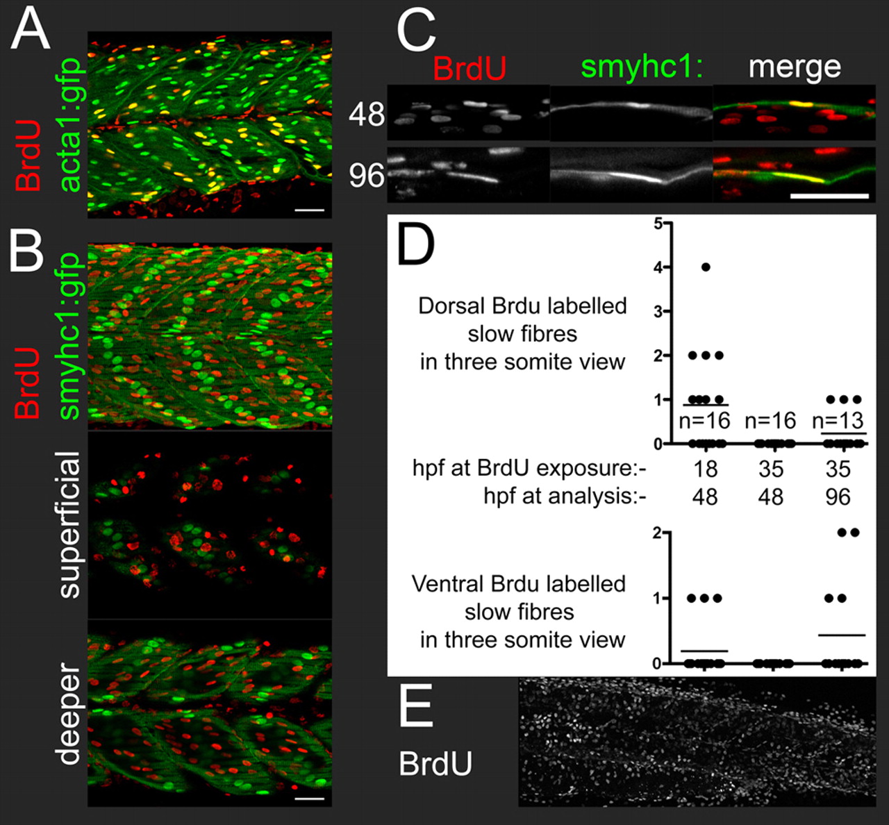
Fig. 4 BrdU labelling shows that wild type embryos have smyhc1-expressing secondary superficial slow fibres. All panels show lateral views of posterior trunk, level with the anus. (A) At 48 hpf, embryos exposed to BrdU at 18 hpf show extensive BrdU co-labelling with acta1:gfp in nuclei that have the characteristic fast muscle elongated and diagonal morphology. (B) At 48 hpf, embryos exposed to BrdU at 18 hpf have BrdU-labelled nuclei superficial to the smyhc1:gfp-labelled surface slow fibres in the presumptive external cell layer (nuclei superficial to the surface slow fibres; these nuclei have a characteristic large flattened disk morphology) and immediately medial to the surface slow fibres, but not in the main body of the surface slow fibre layer (see also Movie 1). (C) BrdU and smyhc1:gfp co-labelling at dorsal somite margin at 48 hpf after BrdU exposure at 18 hpf, and at ventral somite margin 96 hpf after BrdU exposure at 35 hpf. (D) Scatter plots showing number of BrdU and smyhc1:gfp co-labelled nuclei in the dorsal or ventral myotome within a three-somite view at the level of the anus. Each dot represents the count in one embryo. The lines show the mean. Data are shown for exposure to BrdU at 18 hpf or 35 hpf, and with analysis at 48 hpf or 96 hpf. The BrdU labelled smyhc:gfp nuclei were always the smyhc1:gfp nuclei closest to the dorsal or ventral margin of the myotome. (E) At 48 hpf, embryos exposed to BrdU at 35 hpf have extensive labelling in cell types other than the surface slow fibres. Scale bars: 25 μm.