
Image
|
Figure Caption
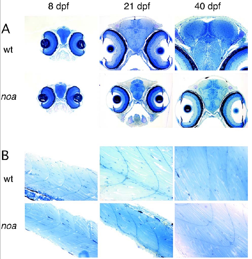
Fig. 10 Histological analysis of noa. (A) Representative brain cross sections of wild-type (wt) and mutant (noa) larvae at 8, 21, and 40 days postfertilization (dpf) shown at x 100 magnification. (B) Representative muscle sagittal sections of wild-type (wt) and mutant (noa) larvae at 8, 21, and 40 dpf shown at x 200 magnification. Fish were fixed and embedded in epon as described (1). Three-micrometer sections were cut on a Leica RM 2155 microtome and stained with methylene blue/azure blue.
Acknowledgments
This image is the copyrighted work of the attributed author or publisher, and
ZFIN has permission only to display this image to its users.
Additional permissions should be obtained from the applicable author or publisher of the image.
Full text @ Proc. Natl. Acad. Sci. USA