
|
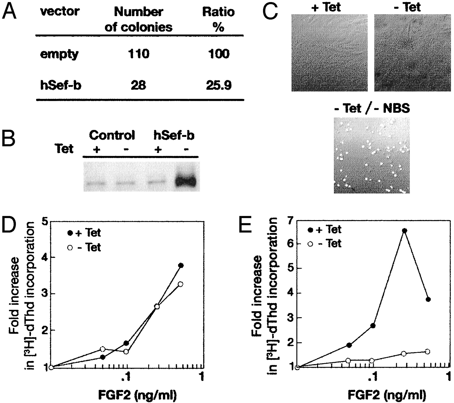
Fig. 3 (A) hSef-b suppresses colony formation in NIH 3T3 cells. Cells were stably transfected with expression vector bearing hSef-b (pCDNA/hSef-b) or an empty vector (pCDNA). After 1 day, cells were diluted (1/25) and marker-selected for 2–3 weeks. Resistant clones were counted at the end of the selection process (five plates for each vector). The results are representative of three experiments. (B) Induced expression of hSef-b in the tet-off NIH 3T3 cells. Cells were grown in 10% serum in the presence and absence of tet. After 24 h, the cells were lysed, and hSef-b expression was analyzed by immunoblotting with hSef-specific antibodies. Control cultures denote parental cells transfected with an empty pTet-splice vector. (C) The effect of hSef-b on apoptosis. NIH 3T3/hSef-b cells were grown for 48 h in the presence or absence of tet or in the absence of tet and serum (–tet, –NBS). Cells were washed and fixed, and apoptosis was then evaluated by terminal deoxynucleotidyltransferase-mediated dUTP nick end labeling. (D and E) hSef-b inhibits the mitogenic activity of FGF2. Confluent cultures of control cells (D) or hSef-b-expressing cells (E) were serum starved and grown in the presence or absence of tet for 24 h. FGF2 was added at the above-indicated concentrations. [3H]Thymidine incorporation assay was performed as described (24, 26).