
|
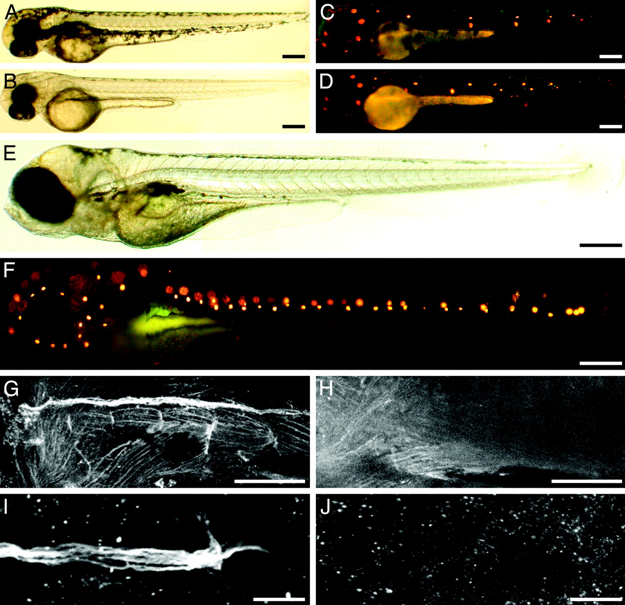
Fig. 4 Additional neuromasts also develop in cls mutant larvae. (A) A bright-field micrograph shows a wild-type larva at 48 hpf. (B) A homozygous cls larva displays a similar pace of development. (C) Exposure of the wild-type animal to the fluorescent compound 4-Di-2-ASP reveals the pattern of functional neuromasts. (D) The mutant larva bears an equivalent number of neuromasts at this stage. (E) A bright-field image of a cls mutant larva at 6 dpf demonstrates the absence of pigmentation on the animal′s head and along its body. (F) Treated with 4-Di-2-ASP, the same animal displays 25 neuromasts on the left side of its trunk. However, the number of neuromasts on the head resembles that of a wild-type larva. (G) The marker 6D2 delineates the distribution of glia in the posterior lateral line of a wild-type larva at 72 hpf. (H) At the same stage, a cls mutant lacks peripheral glia along its trunk. (I) 6D2-positive cells occur along the nerve contacting a neuromast in a wild-type larva. (J) No cells of the posterior lateral-line nerve label with the 6D2 antibody in an ngn1 mutant fish. (Scale bars, 100 μm for A–H and 20 μm for I and J.)