
|
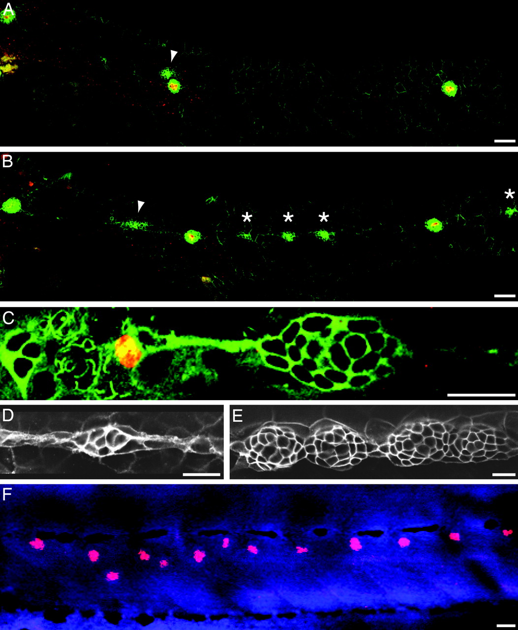
Fig. 3 Interneuromast cells form extra neuromasts in ngn1 mutant larvae. (A) In a wild-type larva at 52 hpf, an antiserum against claudin b (green) reveals cells derived from the posterior lateral-line placode. HCS1 labeling (red) marks mature hair cells; the arrowhead designates the migrating second primordium. (B) At the same stage, a homozygous ngn1 larva displays claudin b-positive cell clusters (asterisks) between the mature neuromasts. The arrowhead marks the second primordium. (C) Labeling of an ngn1 mutant embryo at 52 hpf for phosphohistone H3 (red) shows that interneuromast cells remain mitotically active. Labeling for α-tubulin (green) delineates all cells. (D) Labeling for claudin b demonstrates that the interneuromast cells of an ngn1 mutant begin to coalesce at 52 hpf. (E) By 72 hpf, these cell clusters resolve progressively into new neuromasts. (F)An ngn1 mutant larva labeled for HCS1 (red) and TO-PRO 3 (blue) confirms the presence of mature hair cells within the additional neuromasts at 120 hpf. (Scale bars, 10 μm for C–E and 50 μm for A, B, and F.)