
Image
|
Figure Caption
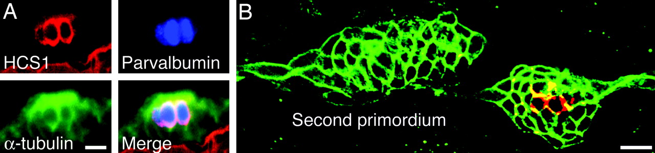
Fig. 2 The HCS1 antiserum marks mature hair cells in neuromasts. (A) In a triply labeled wild-type larva, α-tubulin is present throughout the neuromast, whereas parvalbumin 3 and HCS1 occur in mature hair cells. (B) In a wild-type larva at 48 hpf, a neuromast deposited by the first primordium already bears mature hair cells labeled for HCS1 (red). The migrating second primordium is negative for this marker. Both structures are labeled by immunoreactivity against claudin b (green). (Scale bars, 10 μm.)
Acknowledgments
This image is the copyrighted work of the attributed author or publisher, and
ZFIN has permission only to display this image to its users.
Additional permissions should be obtained from the applicable author or publisher of the image.
Full text @ Proc. Natl. Acad. Sci. USA