
|
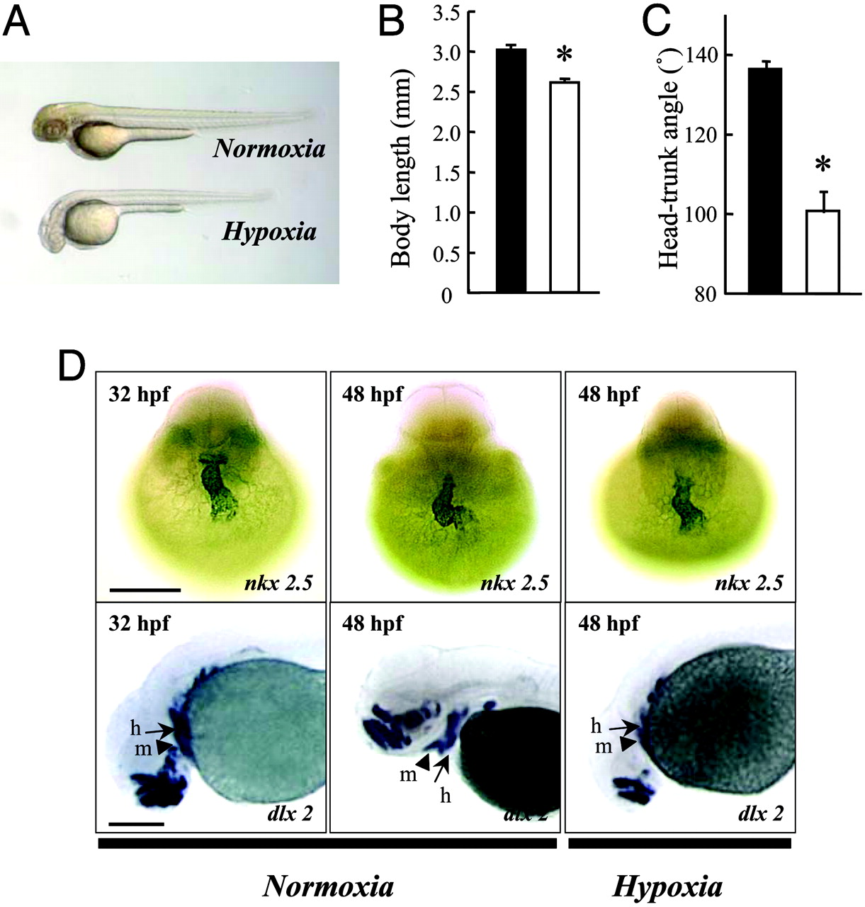
Fig. 1 Hypoxia causes growth retardation and developmental delay. (A) Morphology of wild-type zebrafish embryos at 48 hpf after 24 h of normoxia or hypoxia treatment. (B and C) Effect of hypoxia on embryonic growth and development. Embryos at 24 hpf were transferred to normal (filled bars) or hypoxic (open bars) water. After 24 h, the total body length (B) and HTA (C) were measured. Values are expressed as means ± SE. (n = 28–36). *, P < 0.05. (D) Effect of hypoxia treatment on heart and head skeleton morphogenesis. Embryos raised in water with normal oxygen (normoxia) were fixed at 32 and 48 hpf. A subset of embryos was subjected to 24-h hypoxia exposure beginning at 24 hpf. Whole-mount in situ hybridization was performed by using nkx 2.5 (Upper) and dlx 2 (Lower) riboprobes. h, hyroid arch (arrows); m, mandibular arch (arrow heads). (Scale bar, 200 μm.)