
|
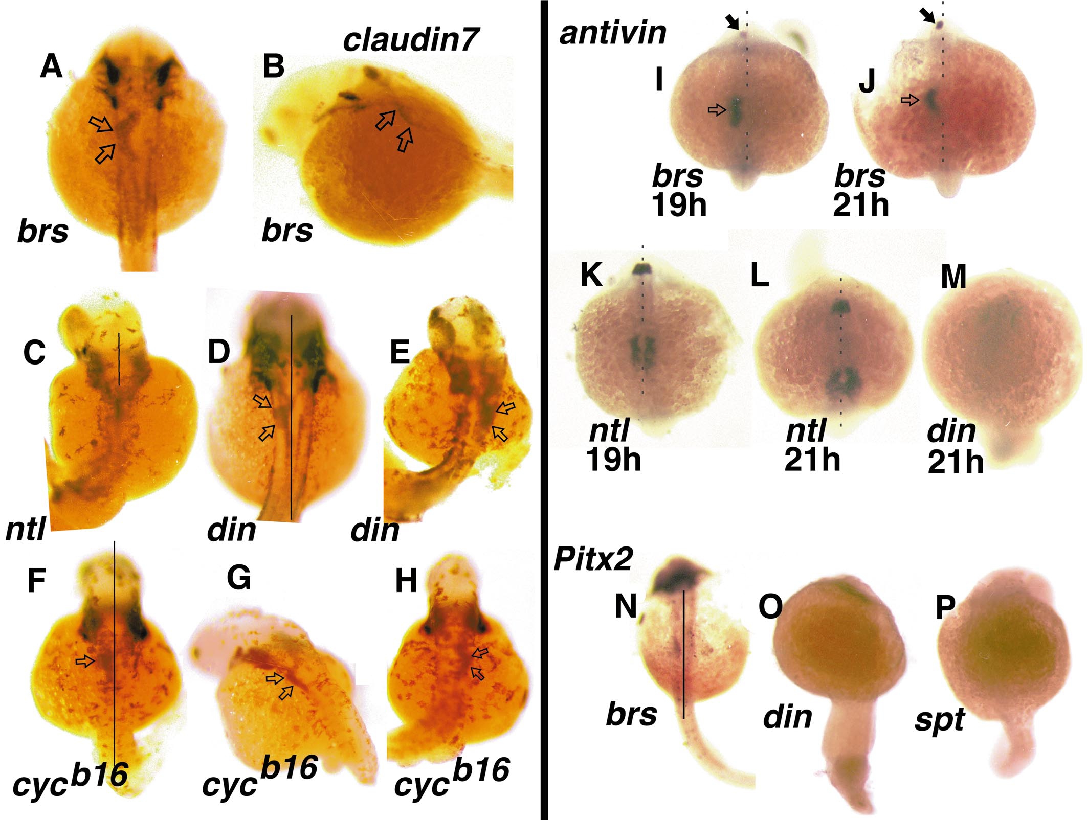
Fig. 7 Zebrafish claudin7 marks the newly looping distal foregut (open arrows), seen in brs embryos viewed dorsally (A) and from a more lateral perspective (B). (C) An example of a ntl homozygote scored while live at the onset of heart contraction as having a left-sided heart; the foregut is midline. din embryos scored as having a left-sided initial heart position display a left-sided foregut (open arrows) at 36 h.p.f. (D) while those with a right-sided initial heart position have a right-sided gut (open arrows) at 36 h.p.f. (E). Finally, similar findings are observed in cycb16 embryos; embryos with left-sided hearts go on to have left-sided foregut (open arrows) seen dorsally (F) and from a posterior oblique view (G), while embryos with right-sided hearts develop a right-sided gut (H). (I–P) Randomization of initial heart tube position is correlated with a failure to correctly express antivin and Pitx2M. antivin is expressed in the left lateral plate mesoderm (open arrow) and left diencephalon (closed arrow) beginning at 19 h.p.f. (I) and continuing at 21 h.p.f. (J) in brs embryos. antivin expression is bilateral in embryos homozygous for ntl (K, L) and absent in embryos homozygous for din (M). Slightly later, Pitx2 is also expressed in left lateral plate mesoderm in brs (N), but not in din (O), or spt (P).
Reprinted from Developmental Biology, 227(2), Chin, A.J., Tsang, M., and Weinberg, E.S., Heart and gut chiralities are controlled independently from initial heart position in the developing zebrafish, 403-421, Copyright (2000) with permission from Elsevier. Full text @ Dev. Biol.