
Image
|
Figure Caption
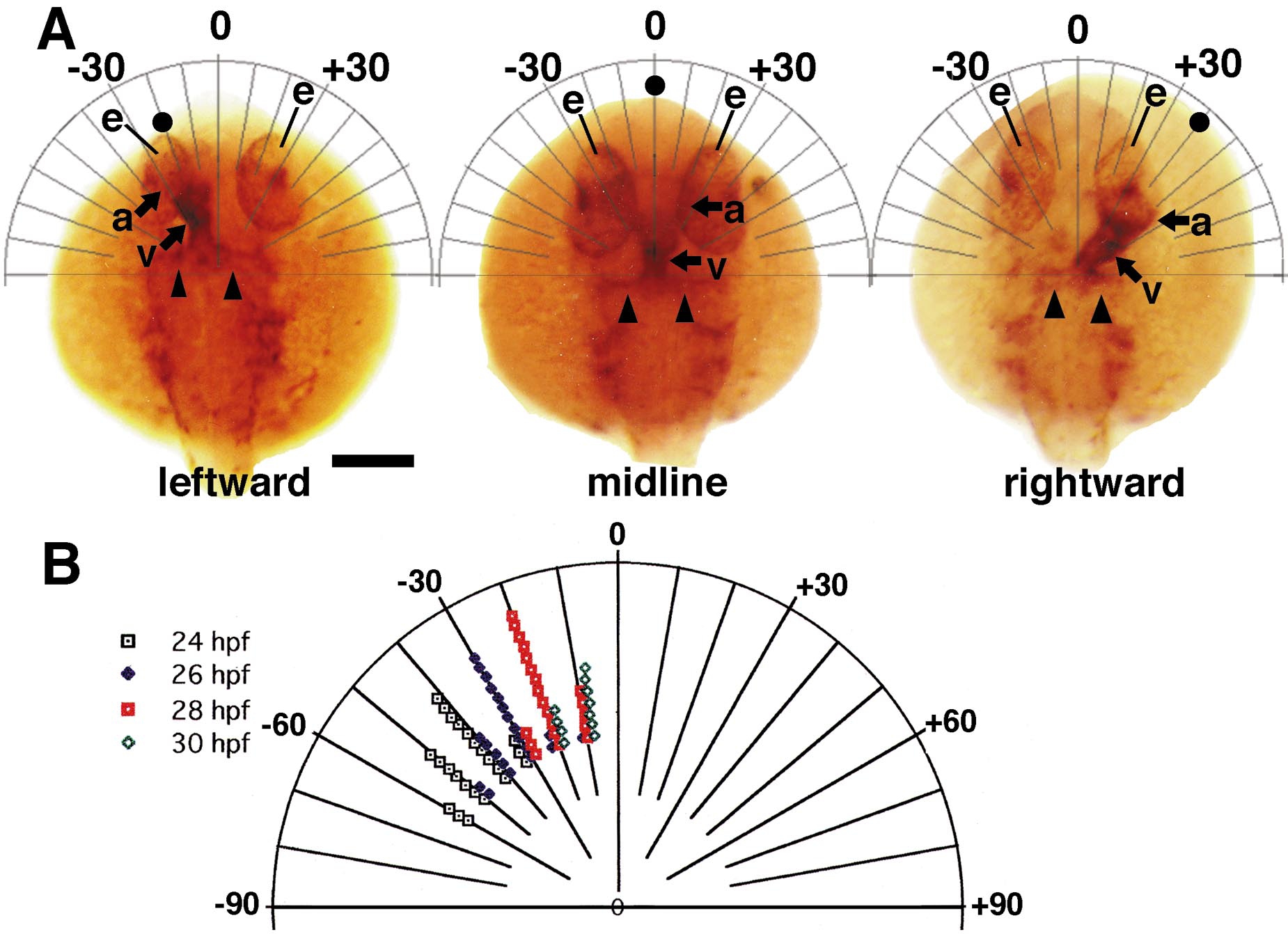
Fig. 2 (A) Method of measuring heart tube angle at 24 h.p.f., illustrated in three flh homozygotes. The examples shown have heart angles of -20°, 0°, and +40°. a, atrium; e, eye; v, ventricle. Arrowheads denote the left and right first aortic arch. Bar 200 μm. (B) Heart tube angle (in degrees) as a function of h.p.f. in brs homozygotes. The embryonic midline is denoted as 0°. The center (origin) of the polar coordinate plot is the site of the caudal end of the heart tube.
Acknowledgments
This image is the copyrighted work of the attributed author or publisher, and
ZFIN has permission only to display this image to its users.
Additional permissions should be obtained from the applicable author or publisher of the image.
Reprinted from Developmental Biology, 227(2), Chin, A.J., Tsang, M., and Weinberg, E.S., Heart and gut chiralities are controlled independently from initial heart position in the developing zebrafish, 403-421, Copyright (2000) with permission from Elsevier. Full text @ Dev. Biol.