
|
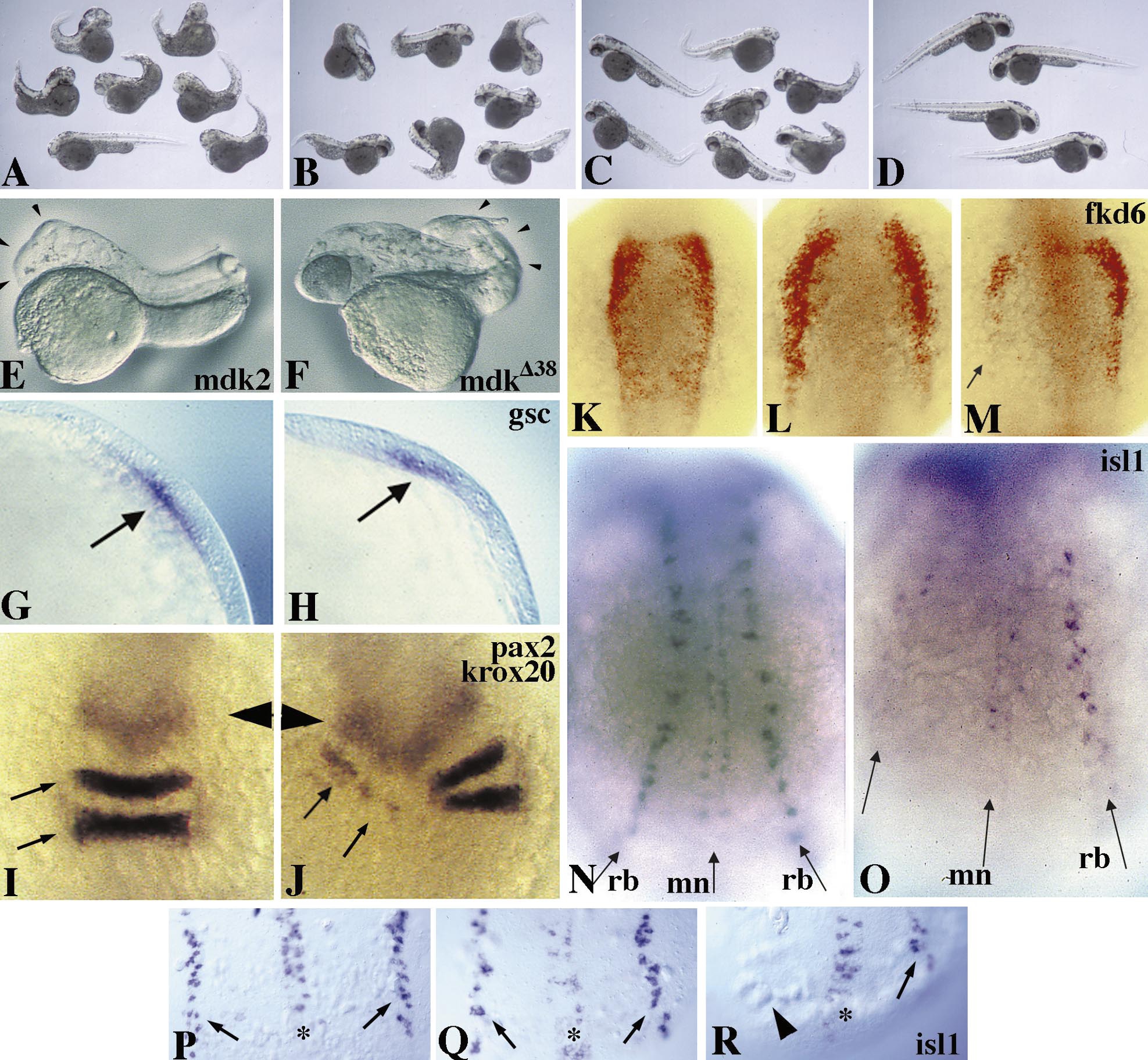
Fig. 4 Repression of posterior neural fates by the dominant interfering mdk2 variant mdkΔ38. (A to D) Coexpression of mdkΔ38 rescues head deficiencies induced by ectopic mdk2 expression. (A) Embryos exhibiting a strong posteriorized phenotype at 48 h after injection with 110 pg mdk2 and 330 pg prolactin RNAs. Note that ectopic mdk2 leads to specific repression of head and eye structures and to some curved tails. (B) Partial rescue of head deficiencies by co-injection of 110 pg mdk2, 220 pg mkΔ38, and 110 pg prolactin RNAs. (C) Greater rescue of head deficiencies by co-injection of 110 pg mdk2 and 330 pg mkΔ38. The majority of head structures appear indistinguishable from those of uninjected control embryos (D). The posterior defects observed are likely due to mdkΔ38 interfering with endogenous mdk2 function. (E, F) 24-h embryos injected with 200 pg wild-type mdk2 (E) and mdkΔ38 (F). Note anterior truncation resulting from gain of function (E, arrowheads) and posterior deficiencies resulting from loss of function (F, arrowheads). (G) Lateral view of gsc expression in an uninjected embryo at 80% epiboly. (H) gsc expression (arrow) is unaffected in mdkΔ38-injected embryos. (I–O) 12-h embryos, dorsal view, anterior to the top. (I) pax2 (arrowhead) and krox20 (arrows) expression in an uninjected embryo. (J) Repression of krox20 expression in rhombomeres 3 and 5 of an embryo unilaterally injected with mdkΔ38. Expression of pax2 at MHB appears normal compared to I. (K) fkd6 staining (red) in an uninjected embryo. (L) Expansion of the fkd6-positive neural crest domain in mdk2-injected embryo. (M) Repression of fkd6 by mdkΔ38 in one half of a unilaterally injected embryo (arrow). (N–R) Ectopic mdkΔ38 expression blocks formation of posterior moto- and sensory neurons. (N) isl1 expression in Rohon–Beard sensory neurons (rb) and motoneurons (mn) of an uninjected embryo at 12 hpf. (O) Unilateral lack of both neural fates in an embryo unilaterally injected with mdkΔ38. (P–R) Dorsal view of 10-h embryos. (P) isl1 expression in an uninjected embryo. (Q) Primary neurons appear unaffected in a mdk2-injected embryo. (R) Note unilateral absence of sensory neurons (arrowhead) in mdkΔ38-injected embryo.
Reprinted from Developmental Biology, 229(1), Winkler, C. and Moon, R.T., Zebrafish mdk2, a novel secreted midkine, participates in posterior neurogenesis, 102-118, Copyright (2001) with permission from Elsevier. Full text @ Dev. Biol.