
|
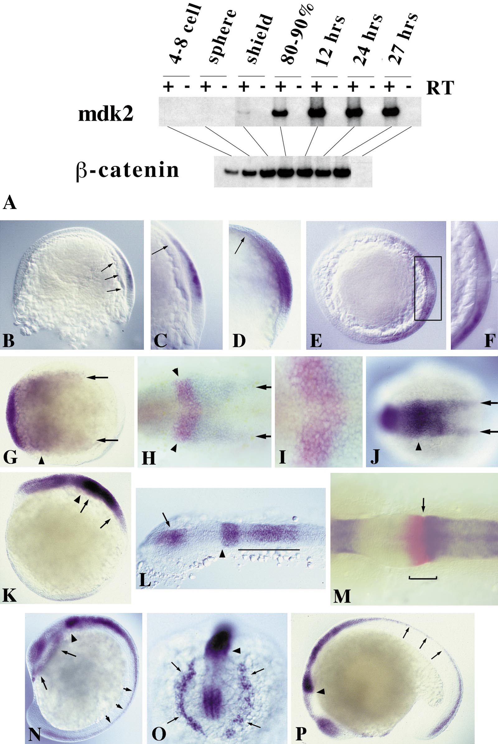
Fig. 2 Expression of mdk2 during zebrafish embryogenesis. (A) RT-PCR of different embryonic stages. β-catenin was used as loading control. (B–N) RNA whole-mount in situ hybridization using full-length mdk2 as probe. (B) 60% epiboly (lateral view, dorsal to the right). Onset of mdk2 expression in the epiblast in a region overlaying the involuting hypoblast (arrows). (C) Close-up of shield region as in B. Arrow indicates anterior border of mdk2 expression overlaying the anterior edge of involuting mesendoderm. (D) 70% epiboly. Expression throughout the presumptive neural plate. Arrow indicates leading edge of axial hypoblast. (E) Optical section through embryo at 70% epiboly at the level of shield region (dorsal to the right). Dorsoventral gradient of mdk2 expression in the epiblast. (F) Higher magnification of shield region (as boxed in E). mdk2 transcripts are excluded from the involuting mesendoderm. (G) 80% epiboly, dorsal view, anterior to the left. Regionalization of mdk2 expression in the early neural plate. Arrowhead indicates elevated expression at the prospective mid–hindbrain boundary (MHB). Arrows demarcate expression at lateral edges of the neural plate. (H) 90% epiboly, dorsal view. Double labeling with eng2 (in red) demarcating future MHB (arrowheads). Arrows indicate mdk2 expression at the edges of the posterior neural plate. (I) Higher magnification of MHB region as in H showing overlapping expression of mdk2 (blue) and eng2 (red). (J) Bud stage, dorsal view. Arrowhead indicates future MHB, arrows denote expression at the edges of the forming neural keel. (K) Lateral view of embryo in J. Arrows mark the developing hindbrain exhibiting a gradient of mdk2 expression with its maximum at the anterior end. Arrowhead indicates strong mdk2 expression at the MHB. (L) 12-h embryo, dorsal view of head region. mdk2 is expressed in the diencephalon (arrow), at the MHB (arrowhead), and in a gradient in the hindbrain, but excluded from the eye fields and midbrain. (M) 12-h embryo, dorsal view of MHB region. Double labeling with eng2 (red). eng2 is expressed across the MHB (bracket), whereas mdk2 marks the anteriormost edge of the metencephalon (arrow). (N) 14-h embryo, lateral view. mdk2 expression at the MHB (arrowhead), in the dorsal neural tube (small arrows), and in dissociating prechordal plate cells underneath the forebrain (large arrows). (O) Frontal view of head region as in N. Arrows indicate dissociating prechordal plate. (P) Latemdk2 expression in fore- and dorsal midbrain, MHB (arrowhead), hindbrain, and dorsal neural tube (arrows).
Reprinted from Developmental Biology, 229(1), Winkler, C. and Moon, R.T., Zebrafish mdk2, a novel secreted midkine, participates in posterior neurogenesis, 102-118, Copyright (2001) with permission from Elsevier. Full text @ Dev. Biol.