
|
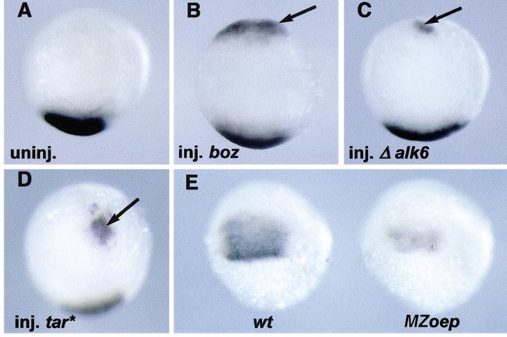
Fig. 10 admp induction at the blastoderm ventral margin. (A–E) In situ hybridization with an admp probe at the shield stage. (A–D) Embryos were injected at the 16-cell stage into one marginal blastomere with nls-gfp RNA as a lineage tracer, sorted according to the position of the injection relative to the dorsal side. Ventrally injected embryos were processed for in situ hybridization (animal pole views, ventral to the top). (A) Uninjected embryo. (B) Ventral injection of boz RNA (2 pg). (C) Ventral injection of Δ alk6 RNA (25 pg). (D) Ventral injection of tar* RNA (2 pg). Arrows indicate the induced admp expression domain. (E) Progeny of an oep-/- X oep-/+ cross. Fifty percent of the embryos (MZoep) had a reduced admp expression domain compared with wild-type siblings (wt).
Reprinted from Developmental Biology, 241(1), Willot, V., Mathieu, J., Lu, Y., Schmid, B., Sidi, S., Yan, Y.-L., Postlethwait, J.H., Mullins, M., Rosa, F., and Peyriéras, N., Cooperative action of ADMP- and BMP-mediated pathways in regulating cell fates in the zebrafish gastrula, 59-78, Copyright (2002) with permission from Elsevier. Full text @ Dev. Biol.