
|
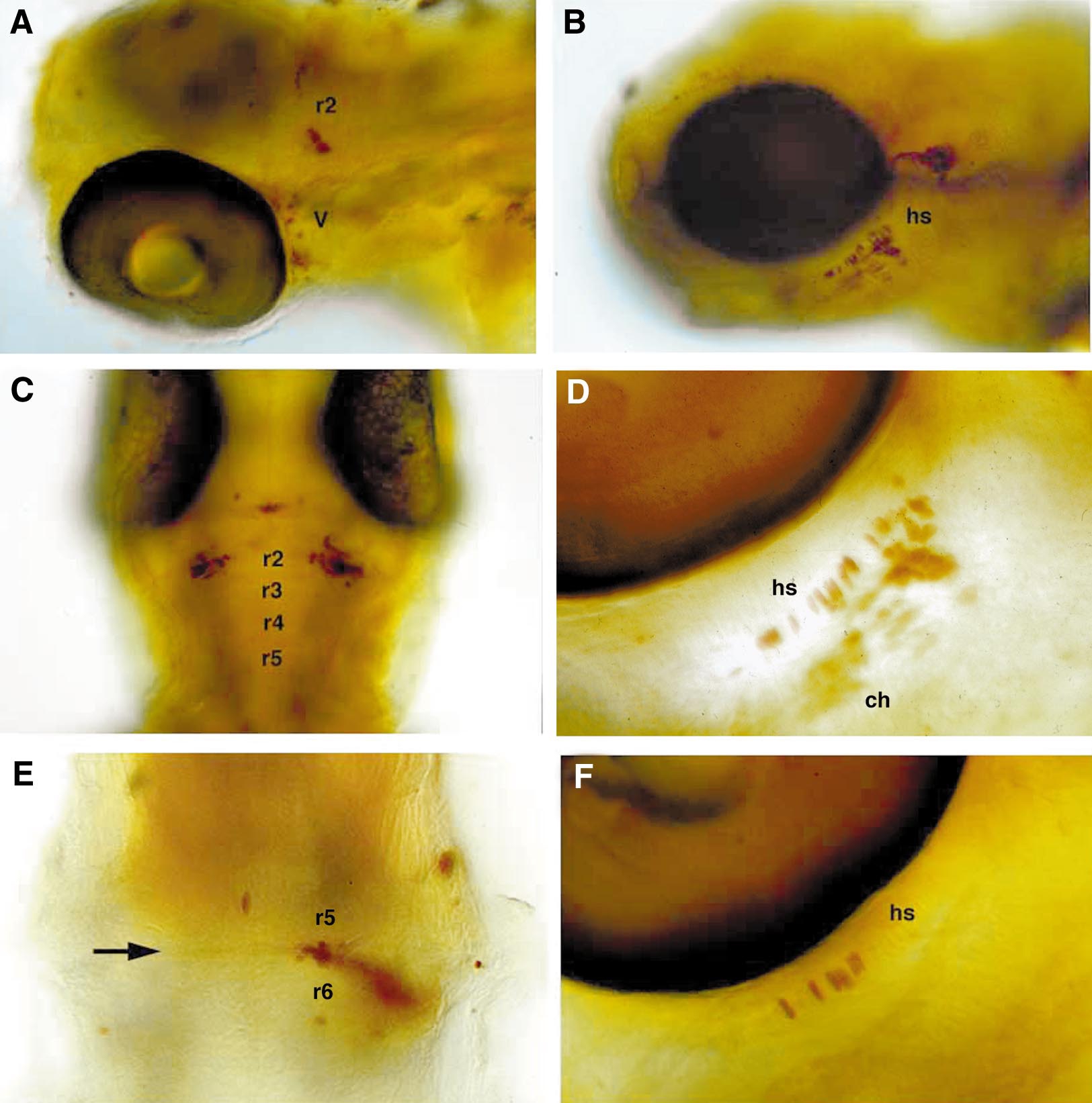
Fig. 9 Cells transplanted along the AP axis regulate their segment-specific skeletal and neural fates. 72-h-old larvae containing transplants were stained for the biotinylated lineage tracer (brown). Left (A, C, E) shows neural derivatives; right (B, D, F) shows skeletal derivatives. (A) Dorsolateral view of cells transposed from r5/6 to r2. Some neural crest cells contained in the same transplant contributed neurons to the trigeminal ganglion of the first arch (V). (B) Lateral view of neural crest transplanted from the r5/6 level to the r3/4 level that contributed to the hyosymplectic cartilage (hs) of the second arch. (C) Dorsal view (anterior to the top) of cells transposed from r5/6 to r2 that contributed bilaterally to r2. (D, F) Ventrolateral views of cells transplanted from r5/6 (D) or r7 (F) that contributed to second arch cartilages. (E) Dorsal view (anterior to the top) of commissural interneurons between r5 and r6, transposed posteriorly from r2/3. Abbreviations: hs, hyosymplectic; ch, ceratohyal; V, trigeminal ganglion.
Reprinted from Developmental Biology, 229, Schilling, T.F., Prince, V., and Ingham, P.W., Plasticity in zebrafish hox expression in the hindbrain and cranial neural crest, 201-216, Copyright (2001) with permission from Elsevier. Full text @ Dev. Biol.