
|
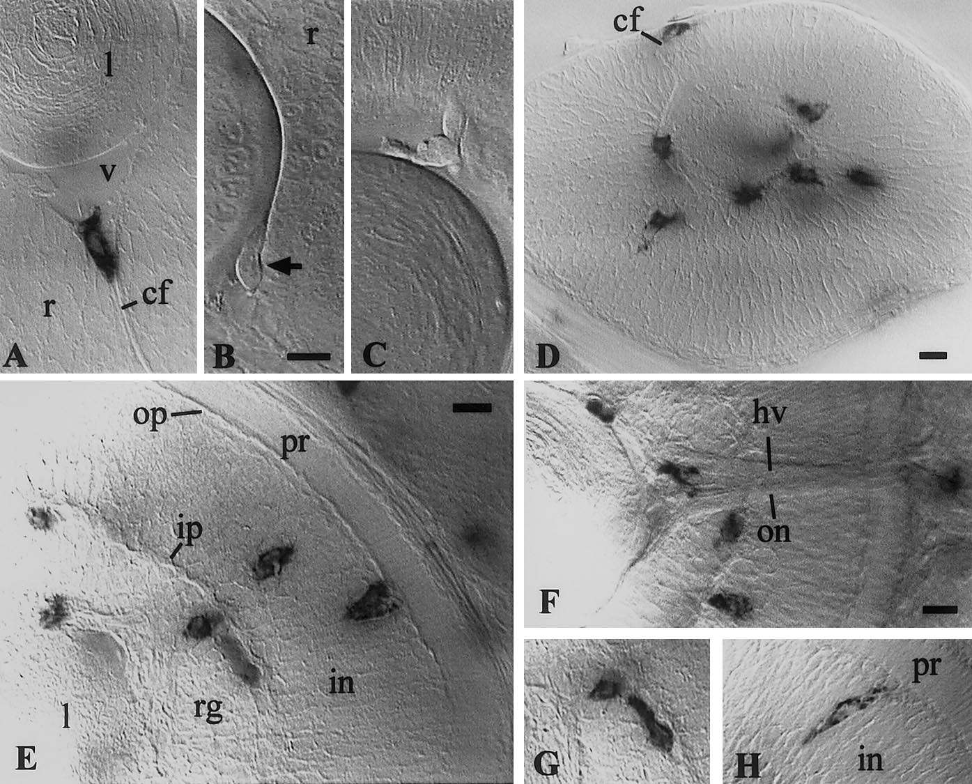
Fig. 5 Early macrophages in the retina. (A, D–H) In situ hybridization for L-plastin. (B, C) Live embryos, 30 hpf. (A, B) macrophage in the choroid fissure, entering the vitreous space, between lens and retina, at 26 (A) and 30 hpf (B). (C) macrophage already in the vitreous space. (D) 35 hpf; macrophages have invaded the vitreous-proximal part of the retina; one is appearing in the choroid fissure. (E–G) 48 hpf; the macrophage lying tangentially along the inner plexiform layer in (E) is shown in focus in (G); (H) 72 hpf. (v) vitreous space; (l) lens; (r) retina; (pr):photoreceptor cell layer; (op) outer plexiform layer; (in) inner nuclear layer; (ip) inner plexiform layer; (rg) retinal ganglion cell layer; (cf) choroid fissure; (on) optic nerve; (hv) hyaloid vein. Bars, 10 μm.
Reprinted from Developmental Biology, 238(2), Herbomel, P., Thisse, B., and Thisse, C., Zebrafish early macrophages colonize cephalic mesenchyme and developing brain, retina, and epidermis through a M-CSF receptor-dependent invasive process, 274-288, Copyright (2001) with permission from Elsevier. Full text @ Dev. Biol.