
Image
|
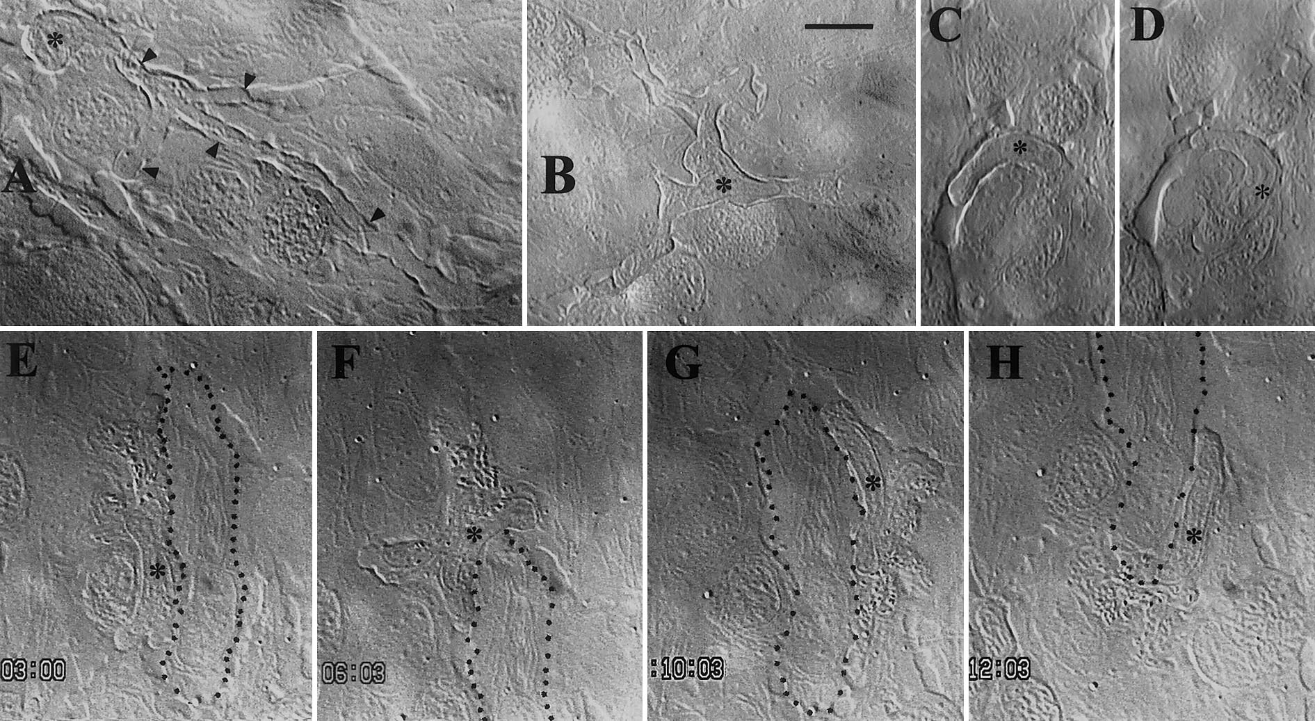
Figure Caption
Fig. 3 Early macrophages in the yolk sac epidermis. Asterisks mark macrophage nuclei. Filaments inside cells are mitochondria. (A, B) 30 hpf; macrophages of the still type, interdigitating (black arrowheads in A) between epidermal cells. (C, D) 30 hpf; macrophage wandering round a small epidermal cell; (D) is 80 s after (C). (E–H) 48 hpf; macrophage wandering all the way round a long epidermal cell (dotted contour) in 12 min. Time indicated in min, s. Bar, 10 μm.
Acknowledgments
This image is the copyrighted work of the attributed author or publisher, and
ZFIN has permission only to display this image to its users.
Additional permissions should be obtained from the applicable author or publisher of the image.
Reprinted from Developmental Biology, 238(2), Herbomel, P., Thisse, B., and Thisse, C., Zebrafish early macrophages colonize cephalic mesenchyme and developing brain, retina, and epidermis through a M-CSF receptor-dependent invasive process, 274-288, Copyright (2001) with permission from Elsevier. Full text @ Dev. Biol.