
|
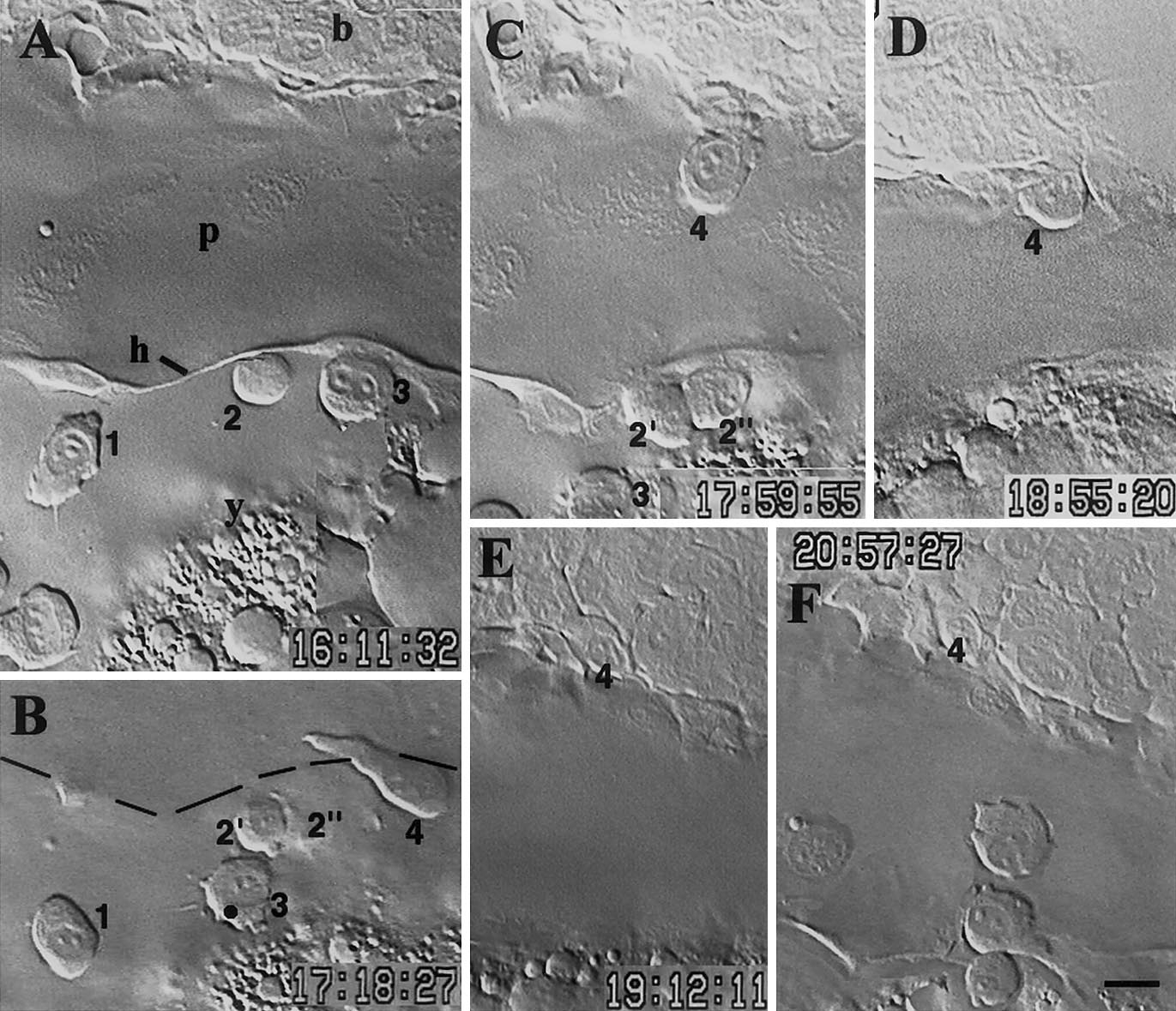
Fig. 1 Early macrophage settling in a branchial arch in the 26- to 30-somite period (22–24 hpf). Sequential lateral views of the same embryo, dorsal upward, rostral to the left. (A) Numbers mark three young macrophages at the lateral border of the pericardial envelope (p), outlined by the pericardial hinge (h, see Fig. 3A in Herbomel et al. 1999). (B) Cell 2 divided; a new young macrophage (4) is climbing on the dorsal side of the pericardium. (C) It now reaches the edge of the branchial arch, (D) progressively enters it, (E) and settles there. (F) Fifteen minutes later, two other young macrophages climbed on the dorsal pericardium by the very same route, but then stayed there, becoming residents of the dorsal pericardium. Time indicated in h, min, s; (b) branchial arches, (y) yolk. Bar, 10 μm.
Reprinted from Developmental Biology, 238(2), Herbomel, P., Thisse, B., and Thisse, C., Zebrafish early macrophages colonize cephalic mesenchyme and developing brain, retina, and epidermis through a M-CSF receptor-dependent invasive process, 274-288, Copyright (2001) with permission from Elsevier. Full text @ Dev. Biol.