
|
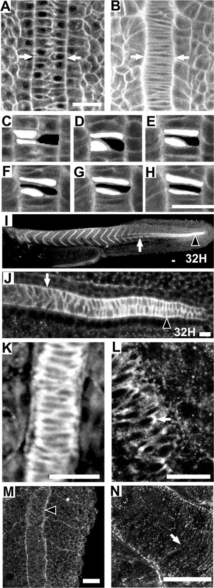
Fig. 5 Zebrafish notochord cells intercalate as in Xenopus embryos; Fak protein is highly expressed in the region of the notochord that is undergoing convergence; and Fak is phosphorylated at the notochord-somite boundary. Anterior is towards the top or left in all panels. Dorsal views except (I) and (J), which are side views. (A–H) Time-lapse analysis of a vitally-stained zebrafish embryo (see methods) from the 1-somite stage through the 4-somite stage at the level of the first 4 somites. At time 0 min (A), the notochord is two cells wide (white arrows) and by time 38 min (B) cellular intercalation results in a notochord that is one cell wide. (C–H) Enlarged view of three cells intercalating. (C) 0 min. (D) 10 min. (E) 23 min. (F) 30 min. (G) 35 min. (H) 40 min. (I–L) Immunostaining for Fak protein. The posterior region of the notochord undergoing cell intercalation is noted with an arrowhead. White arrow indicates the notochord–somite boundary anterior to the intercalating domain. (K) A 10-somite embryo, dorsal view at the level of somite 4 showing the notochord–somite boundary. (L) Enlarged view of a notochord undergoing intercalation in a 10-somite embryo, at the level of somite 8. An arrow notes a discrete plaque of Fak. (M, N) Immunostaining for pY397FAK. M: A 13-somite embryo at somite 13 stained for phospho-Fak at the notochord-somite boundary (black arrowhead). (N) Projection of confocal sections that include the entire thickness of notochord of a 13-somite embryo showing circumferential striations in phospho-Fak staining perpendicular to the notochord axis. Scale bars, 20 μm, with the exception of (I), where scale bar is 40 μm.
Reprinted from Developmental Biology, 240(2), Henry, C.A., Crawford, B.D., Yan, Y.-L., Postlethwait, J., Cooper, M.S., and Hille, M.B., Roles for zebrafish focal adhesion kinase in notochord and somite morphogenesis, 474-487, Copyright (2001) with permission from Elsevier. Full text @ Dev. Biol.