
Image
|
Figure Caption
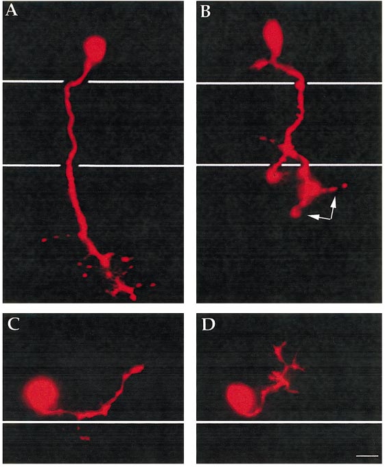
Fig. 4 CaP and MiP motor axons display abnormalities in desb420 mutants. Lateral view of pseudocolor images of live CaP (A, B) and MiP (C, D) motoneurons labeled with rhodamine dextran and visualized at approximately 26 h in wild-type (A, C) and desb420 mutant (B, D) embryos. Cell bodies reside in the spinal cord and white lines denote the dorsal and ventral aspects of the notochord (A, B) and the dorsal aspect of the notochord (C, D). Scale bar, 10 μm.
Figure Data
Acknowledgments
This image is the copyrighted work of the attributed author or publisher, and
ZFIN has permission only to display this image to its users.
Additional permissions should be obtained from the applicable author or publisher of the image.
Reprinted from Developmental Biology, 237(2), Gray, M., Moens, C.B., Amacher, S.L., Eisen, J.S., and Beattie, C.E., Zebrafish deadly seven functions in neurogenesis, 306-323, Copyright (2001) with permission from Elsevier. Full text @ Dev. Biol.