
|
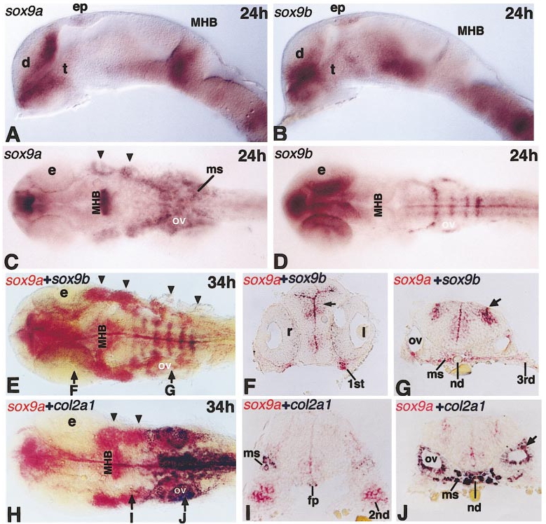
Fig. 6 Expression patterns of sox9a and sox9b in the head during the pharyngula stage. (A, B) Lateral view and (C, D) dorsal view of the 24 h head. In (A) and (B), both sox9a and sox9b transcripts were detected in the epiphysis (ep), forebrain, and the ventral midbrain. Expression of sox9a in the diencephalon (d) of the forebrain extends dorsally, whereas sox9b expression is mainly at the ventral diencephalon. t, telencephalon. Only sox9a transcripts are detected in the midbrain-hindbrain boundary (MHB), pharyngeal pouches (arrowheads in C), and the head mesenchyme (ms) in the caudal hindbrain. Transcripts of sox9b are detected in the eye (e) close to the midline and in six stripes of the hindbrain, with the highest expression in the four posterior rhombomeres (D). Otic vesicle (ov) marks the position of the caudal hindbrain. (E–G). Double staining with sox9a (red) and sox9b (blue) at 34 h. Both whole mount (E) and cryosections (F, G) show that both genes are expressed in cells around the ventricle (arrow in F) and in two areas (arrow in G) in the dorsal hindbrain. In the head mesenchyme (ms) and segmented pharyngeal arch primordia, which are shown in the whole mount (as arrowheads in E) or in sections (labeled as 1st, 2nd, 3rd), only sox9a transcripts were detected. As in panel D, the retina (r), but not the lens (l) of the eye (e), retains sox9b expression. (H–J) Double staining with sox9a (red) and col2a1 (blue) of the head. The precondensed mesenchyme in the segmented pharyngeal arches express only sox9a, not col2a1 (arrowheads in H and 2nd in I). Transverse sections through the rostral hindbrain (I) and at the level of the ear (J) confirm that the transcripts of the two genes overlap in the head mesenchyme (ms) which lie beneath or to the side of the neural tube. Expression of col2a1 in the epithelium (ep) of otic vesicle is much stronger than that of sox9a. Only a few epithelial cells are positive for both sox9a and col2a1 staining (arrow in J). Expression of col2a1 was also detected in the midline structures, including the floor plate (fp) and notochord (nd), but not in CNS.
Reprinted from Developmental Biology, 229, Chiang, E., Pai, C.-I., Wyatt, M., Yan, Y-L., Postlethwait, J., and Chung, B.-C., Two sox9 genes on duplicated zebrafish chromosomes: expression of similar transcription activators in distinct sites, 149-163, Copyright (2001) with permission from Elsevier. Full text @ Dev. Biol.