
|
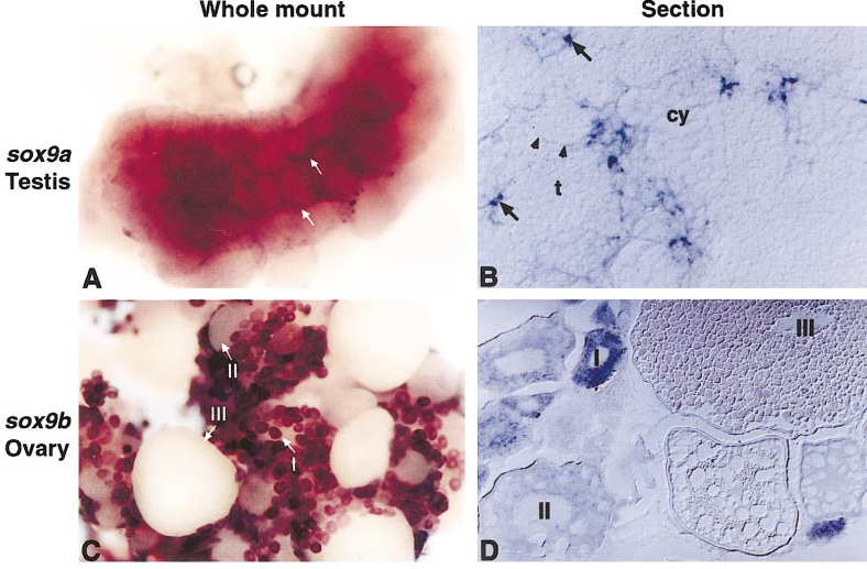
Fig. 5 In situ hybridization shows transcripts of sox9a in zebrafish testis and sox9b in the ovary. (A, C) Whole-mount (B, D) sections of testis and ovary. Transcripts of sox9a (A, B) were detected in the zebrafish testis. Scattered cells across the mature testis express sox9a (arrows in A and B). Cryosection revealed that these cells are Sertoli cells (arrows B), which are irregularly shaped and lie close to the edge of the large membrane-bound cyst-like structures in the testis. Spermatogenesis proceeds synchronously within each cyst. Different stages of spermatogenesis can be seen in different cysts. Large and round shaped germ cells are early stage spermatocytes (cy). These spermatocytes decrease in size as spermatogenesis progresses and develop into spermatids (t). Arrowheads in B indicate the edge of one of the cysts. Transcripts of sox9b (C, D) were evident in the previtellogenic oocytes of the ovary. I, follicles at the primary growth stage (stage I); II, follicles at the cortical alveolus stage (stage II); III, stage III vitellogenic follicles.
Reprinted from Developmental Biology, 229, Chiang, E., Pai, C.-I., Wyatt, M., Yan, Y-L., Postlethwait, J., and Chung, B.-C., Two sox9 genes on duplicated zebrafish chromosomes: expression of similar transcription activators in distinct sites, 149-163, Copyright (2001) with permission from Elsevier. Full text @ Dev. Biol.