
|
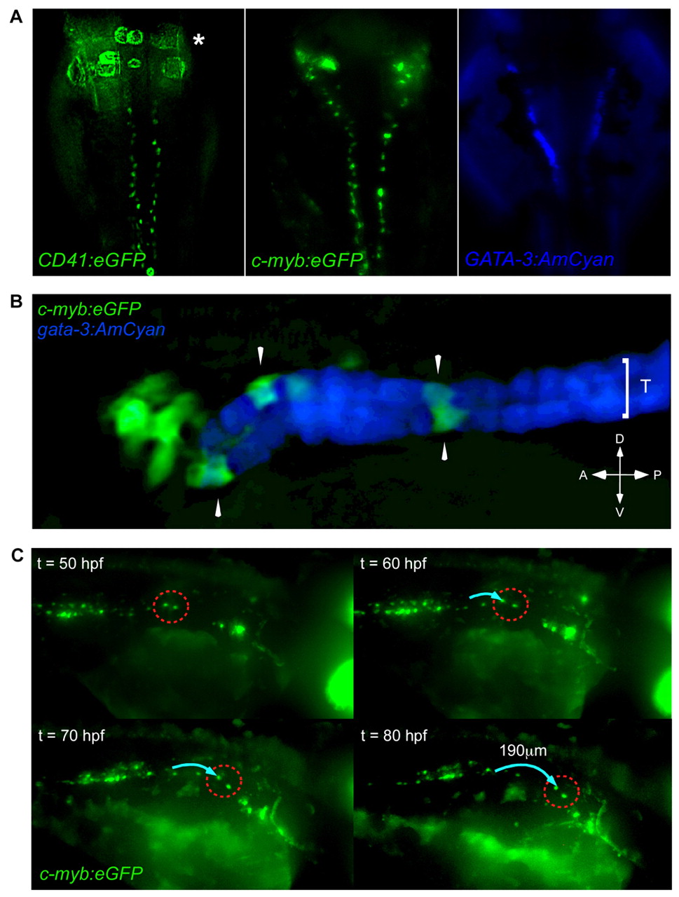
Fig. 6 Hematopoietic precursors migrate along the pronephric tubules. (A) CD41:eGFP (left panel; asterisk marks fluorescence from circulating thrombocytes) and cmyb:eGFP (middle panel) transgenes are expressed in cells along each pronephric tubule. The anterior pronephric tubules are marked by a gata3:AmCyan transgene (right panel). Dorsal views of animals with anterior side upwards. (B) cmyb:eGFP+ cells (arrowheads) are localized upon gata-3:AmCyan+ pronephric tubules (T). (C) Timelapse imaging demonstrates cmyb:eGFP+ cells migrate along the pronephric tubules in an anterior direction. Two GFP+ cells (dotted red circle) were observed to migrate ∼190 μm (blue arrow) over 30 hours. Embryos imaged dorsal side upwards, anterior towards the right.