
|
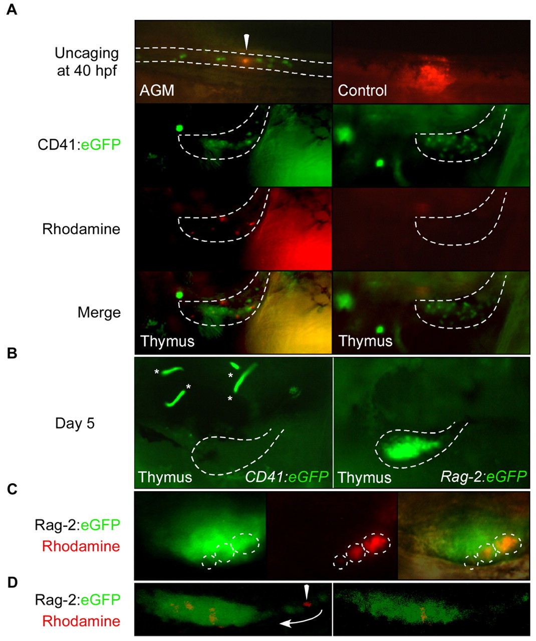
Fig. 4 AGM CD41:eGFP+ cells seed the thymus to become rag2+ thymocytes. (A) Upper left panel shows one CD41:eGFP+ cell immediately after rhodamine uncaging at 40 hpf (arrowhead). Ten cells were uncaged per embryo, and thymic lobes (areas within broken lines in lower panels) were analyzed at 4 dpf. Rhodamine+ cells were observed in the thymic lobes, along with GFP+ cells that were not uncaged (lower left panel). Control animals where regions outside of the AGM were laser uncaged never showed rhodamine+ thymic cells (right panels). (B-D) Similar uncaging experiments using CD41:eGFP, Rag-2:eGFP double transgenic animals show labeled thymic immigrants are lymphoid. (B) CD41:eGFP+ cells were laser targeted at 40 hpf in the AGM and thymi analyzed at 5 dpf, when thymic cells no longer express the CD41 transgene (left panel; asterisks mark circulating CD41+ thrombocytes) and when nascent thymocytes robustly express the rag2 transgene (right panel). (C) Targeted CD41:eGFP+ cells migrate to the thymus and express the rag2 transgene. Left panel shows GFP expression in a representative thymic lobe, middle panel clones of rhodamine+ cells and right panel a merged imaged, including Nomarski overlay. (D) Confocal imaging of targeted thymic immigrants. Left panel shows a maximum projection of the entire thymic lobe, and shows a rhodamine+ GFP- cell migrating (arrowhead) into the thymus via a posterior thymic duct (arrow). Right panel shows a single z-slice through the thymus showing expression of GFP and rhodamine. All embryos oriented dorsal side upwards, anterior towards the left.