
|
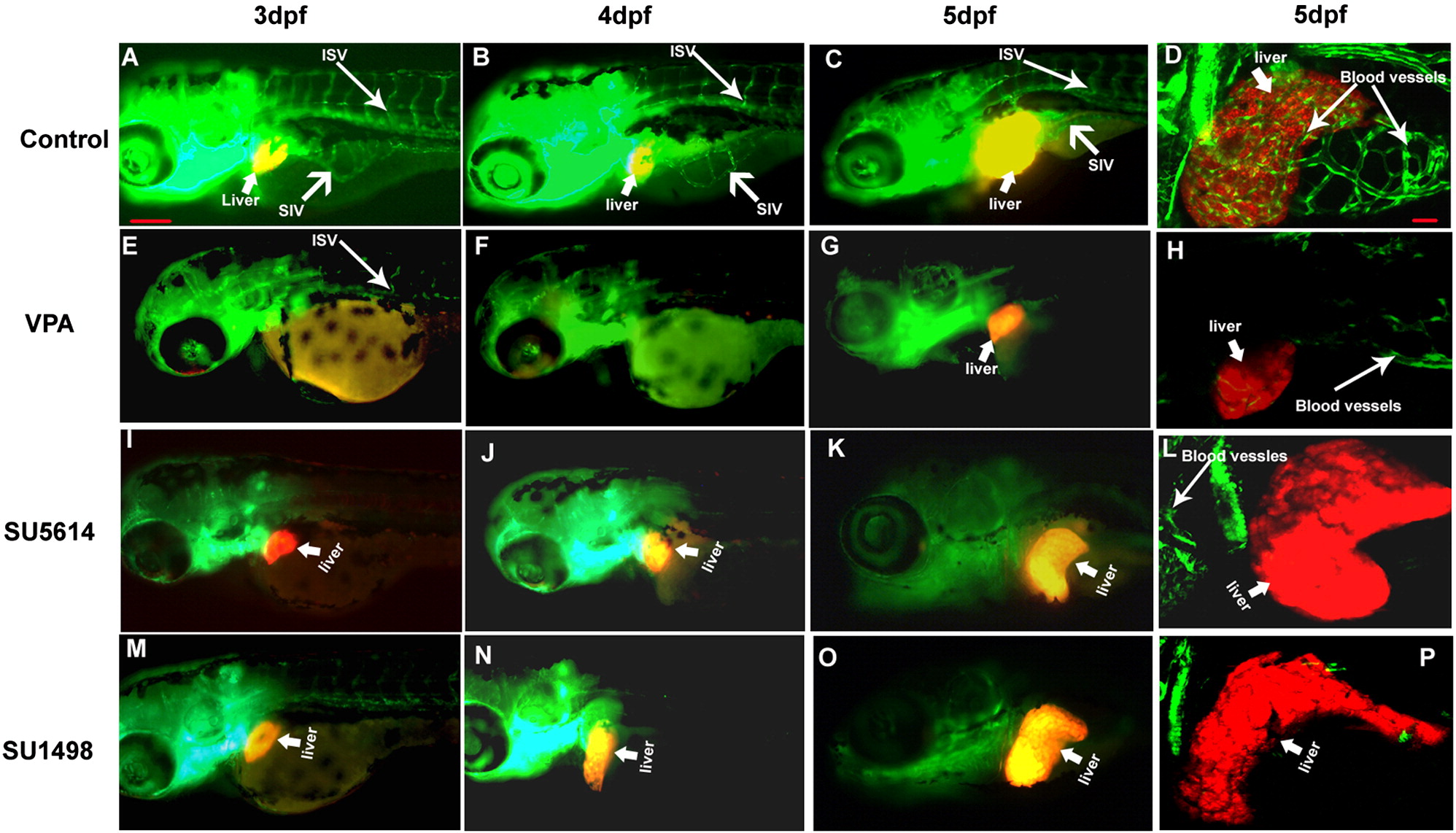
Fig. 3 Liver formation in zebrafish does not require vascularization. Zebrafish embryos from triple transgenic line were treated with VPA (20 μM). Severe defects in intersomatic blood vessels (ISV) (thin white arrows) and subintestinal blood vessels (SIV) (wide white arrow) were observed up to 5 dpf (E–H). Liver only appeared on 5 dpf and no or minimum liver vascularization was observed (H). Similar angiogenesis defects were observed in embryos treated with angiogenesis inhibitors SU5614 (5 μM, I–L) and SU1498 (5 μM, M–P). However, liver (thick white arrows) formed normally on 3 dpf and grew extensively from 3 dpf to 5dpf in these embryos (I–P). All images are lateral view, anterior toward left. The images D, H, L and P are confocal images of merged z-stacks. The scale bar is 100 μm in panel A, and 30 μm in panel D.
Reprinted from Developmental Biology, 317(1), Farooq, M., Sulochana, K.N., Pan, X., To, J., Sheng, D., Gong, Z., and Ge, R., Histone deacetylase 3 (hdac3) is specifically required for liver development in zebrafish, 336-353, Copyright (2008) with permission from Elsevier. Full text @ Dev. Biol.