
Fig. 5
- ID
- ZDB-IMAGE-080501-14
- Publication
- Zeller et al., 2002 - Migration of zebrafish spinal motor nerves into the periphery requires multiple myotome-derived cues
- All Figures
- Figures for Zeller et al., 2002
|
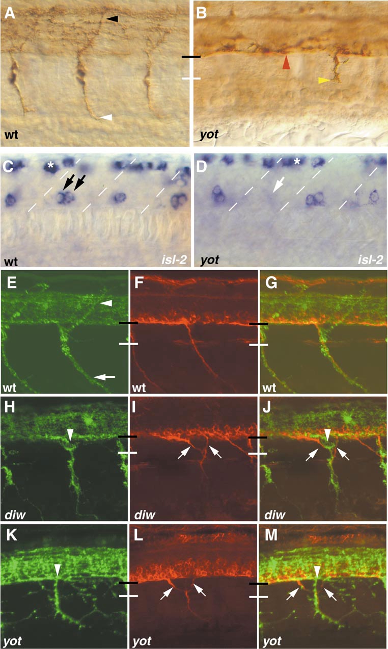
Fig. 5 In diwanka and you-too/gli2 mutants, pathfinding errors of secondary motoneurons occur independent of primary motor axons. Lateral views of a wild-type (A) and a you-too/gli2 mutant (B) embryo at 23 hpf stained with znp-1 antibody. (A) In each wild-type hemisegment, a CaP (white arrowhead) and a MiP axon (black arrowhead) are present. (B) In you-too/gli2 embryos, primary motor axons migrate within the spinal cord (red arrowhead), and in only 20% of the hemisegments, motor axons exit the spinal cord but stall at the distal end of the common path (yellow arrowhead). In over 80% of the hemisegments, primary motor axons completely fail to exit into the periphery (n = 100). (C, D) In the ventral spinal cord islet-2 labels CaP and VaP primary motor neurons (black arrows; VaP neurons are present in only 50% of wild-type hemisegments). In you-too/gli2 embryos (D), there is a slight reduction in the number of primary motor neurons (white arrow). Asterisks indicate islet-2-positive sensory neurons in the dorsal spinal cord. (E-M) Confocal analysis of double labeled motor neurons using znp-1 (green, E, H, K) and zn-5 (red, F, I, L) in 38-hpf wild-type (E–G), diwanka (H–J), and you-too/gli2 embryos (K–M). (G, J, M) Both channels. (E–G) In wild-type embryos, znp-1 labels primary and secondary motor axons extending along the common path (between black and white bars), and along the ventral path (white arrow). Note that the dorsal path (white arrowhead) is znp-1-positive, but zn-5-negative, indicating that at 38 hpf, fasciculated secondary motor axons have not extended along the dorsal path. (H–J) In diwanka mutant embryos, zn5-positive motor axons exit ectopically from the spinal cord (arrows) ignoring the path of pioneering znp-1-positive primary motor axons (arrowhead). (K–M) you-too/gli2 embryos display similar phenotypes, demonstrating that, unlike wild-type axons, mutant secondary motor axons (zn5-positive, in red) can ignore pioneering primary motor axons.
Reprinted from Developmental Biology, 252(2), Zeller, J., Schneider, V., Malayaman, S., Higashijima, S., Okamoto, H., Gui, J., Lin, S., and Granato, M., Migration of zebrafish spinal motor nerves into the periphery requires multiple myotome-derived cues, 241-256, Copyright (2002) with permission from Elsevier. Full text @ Dev. Biol.