
Fig. 3
- ID
- ZDB-IMAGE-080501-10
- Publication
- Zeller et al., 2002 - Migration of zebrafish spinal motor nerves into the periphery requires multiple myotome-derived cues
- All Figures
- Figures for Zeller et al., 2002
|
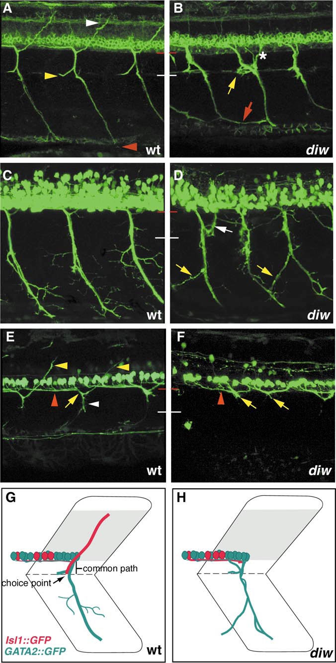
Fig. 3 Pathfinding defects in diwanka mutant embryos. In 52-hpf wild-type embryos (A), secondary motor axons have completed migration along the common path (between red and white line) and extend on their cell-type-specific paths to the dorsal (white arrowhead), medial (yellow arrowhead), and ventral myotome (red arrowhead), respectively. In diwanka mutant embryos (B), zn-5 staining reveals multiple pathfinding defects. Motor axons exited the spinal cord within two independent segmental nerves (asterisk), ventral nerves appeared thinner and less fasciculated and often projected aberrantly into neighboring hemisegments (red arrow). Rostral nerves were absent or less fasiculated (yellow arrow), while projections into the dorsal myotome were absent in all hemisegments. In wild-type embryos, TG(GATA2:GFP)-expressing motor nerves have completed migration along the common path and extended on their cell-type specific paths into the ventral myotome (C). The ventral nerve has formed secondary and tertiary branches that do not cross segmental boundaries. The white line indicates the distal end of the common path. In diwanka mutant embryos (D), TG(GATA2:GFP)-expressing secondary motor axons exit the cord and project though multiple segmental nerves (white arrow), appear thinner and less fasciculated and invade adjacent somites (yellow arrows). In wild-type embryos carrying the TG(ISL1:GFP) transgene (E), labeled motor axons can be detected within the spinal cord extending towards the segmental exit point (red arrowhead), along the common path (white arrowhead), and along their cell-type-specific path into the dorsal myotome (yellow arrowhead). In diwanka mutant embryos expressing TG(ISL1:GFP) (F), secondary motor axons extend inside the spinal cord (red arrowhead) but completely fail to enter the common path. Instead their growth cones stall and accumulate at the segmental exit point (yellow arrows). Schematic summary of wild-type trajectories (G) and diwanka pathfinding defects (H) using TG(GATA2:GFP) (green) and TG(ISL1:GFP) transgenic lines.
Reprinted from Developmental Biology, 252(2), Zeller, J., Schneider, V., Malayaman, S., Higashijima, S., Okamoto, H., Gui, J., Lin, S., and Granato, M., Migration of zebrafish spinal motor nerves into the periphery requires multiple myotome-derived cues, 241-256, Copyright (2002) with permission from Elsevier. Full text @ Dev. Biol.