
|
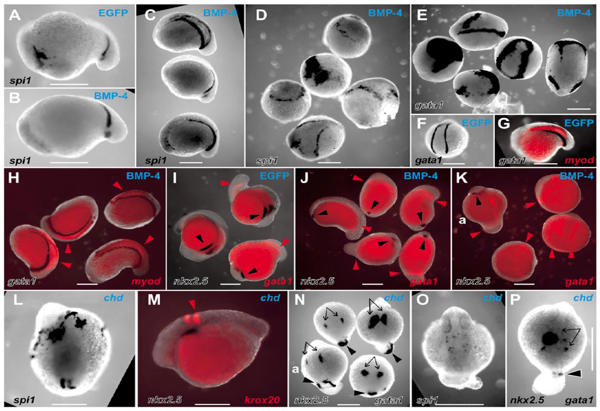
Fig. 8 Actions of BMP and chordin on the LPM to perturb the balance of rostrocaudal LPM fates. Whole-mount in situ hybridization preparations of zebrafish embryos displaying expression domains of various markers (indicated in lower lefthand corner of each panel), in EGFP mRNA-injected (A, F, G, I), Xenopus BMP-4 mRNA-injected (B–E, H, J, K) and zebrafish chordin mRNA-injected (L–P) embryos (injected mRNA indicated in top righthand corner of each panel). Embryos are variously orientated to optimally display the range of expression patterns of interest. (A–N) Embryos are at approximately 14 hpf; (O, P) Approximately 24 hpf stages of development. (A–D) spi1 expression in control EGFP-injected (A), moderately (B and C), and severely (D) affected BMP-4-injected embryos. (A–C, approximately lateral views with head to the left; the five dysmorphic embryos in D are in various orientations.) (E, F) gata1 expression in five severely affected BMP-4-injected embryos (E, various orientations), showing ectopic expression and expansion of gata1 expression compared with the wild-type expression pattern observed in EGFP-injected embryos (F). (G, H) Group of four moderately BMP-4-affected embryos (H) displaying normal expression of gata1 and myod (red arrowheads) (various orientations), compared with the wild-type expression pattern observed in EGFP-injected embryos (G). (I, J) Group of five moderately BMP-4-affected embryos (J) showing normal gata1 expression (red arrowheads) but markedly diminished nkx2.5 expression (black arrowheads) (various orientations), compared with the wild-type expression pattern observed in EGFP-injected embryos (I). (K) Three severely affected BMP-4-injected embryos showing expanded gata1 expression and loss of nkx2.5 expression, including a relatively unaffected embryo (upper left, marked “a”) for comparison with rostral nkx2.5 strips (black arrowhead) and caudal gata1 stripes (red arrowheads). (L) Dorsal view (head at top) of a chordin-injected embryo showing extensive rostral spi1 and diminished caudal spi1 expression domains. (M) Lateral view (head to left) showing relationship of nkx2.5 and krox20 in moderately chordin-affected embryo. (N) Dorsal views (head to top) of three moderately chordin-affected embryos showing markedly reduced extent of caudal gata1 expression (black arrowheads) and less affected more rostral nkx2.5 expression (black arrows), with a minimally affected embryo (lower left, marked “a”) for comparison. The wild-type expression pattern of these genes in EGFP-injected embryos is shown in two colors (G, I). (O, P) Dorsal views (head to top) of moderately affected chordin-injected embryos showing preserved dispersed spi1 expression (O), bilateral nkx2.5 expression (P, arrows), and small spot of gata1 expression in tail stub (P, arrowhead). Images are representative of the spectrum of effects observed in 137 BMP-4- and 78 zebrafish chordin-injected embryos scored at ≈14 hpf (19/137 and 41/78 severely affected, respectively), and 154 BMP-4- and 66 chordin-injected embryos scored at ≈24 hpf (21/154 and 13/66 severely affected, respectively), collected from several independent injection days (BMP-4, n = 3; chordin, n = 2) and divided between the various groups of in situ hybridization analyses. In these experiments, control embryos were injected with 10 pg of mRNA encoding EGFP with a <5% abnormality rate (94 embryos, which were divided into groups to demonstrate the wild-type pattern for the various in situ hybridization expression analyses). Scale bars, 0.3 mm.
Reprinted from Developmental Biology, 246(2), Lieschke, G.J., Oates, A.C., Paw, B.H., Thompson, M.A., Hall, N.E., Ward, A.C., Ho, R.K., Zon, L.I., and Layton, J.E., Zebrafish SPI-1 (PU.1) marks a site of myeloid development independent of primitive erythropoiesis: implications for axial patterning, 274-295, Copyright (2002) with permission from Elsevier. Full text @ Dev. Biol.