
|
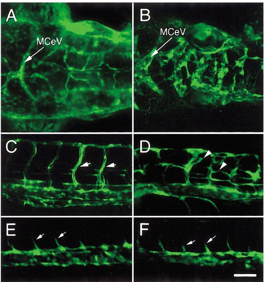
Fig. 6 Vascular defects in TG(fli1:EGFP)y1; mibta52b embryos. (A) Head of a wild type TG(fli1:EGFP)y1 embryo at 2 dpf. Arrow indicates midcerebral vein which runs along the midbrain–hindrain boundary. (B) Head vasculature within the hindbrain (posterior to the midcerebral vein) of a TG(fli1:EGFP)y1; mibta52b mutant embryo is severely disorganized and highly dilated. The location and pattern of the midcerebral vein appears normal, although it appears dilated. (A, B) Dorsal views, anterior is to the left. (C) Segmental vessels (arrows) appear at regular intervals corresponding to the vertical myosepta of the somites in a wild type TG(fli1:EGFP)y1 embryo. (D) In TG(fli1:EGFP)y1 embryos mutant for mibta52b, ectopic segmental vessel sprouts (arrowheads) are seen throughout the trunk. (E) A wild type sibling TG(fli1:EGFP)y1 at the 20-somite stage with segmental vessels (arrows) sprouting from the dorsal aorta. (F) Segmental vessel sprouts appear normal inTG(fli1:EGFP)y1; mibta52b mutant embryos at the 20-somite stage. (C–F) Lateral views, anterior is to the left, dorsal is up. Scale bar, (C–F) 50 μm.
Reprinted from Developmental Biology, 248(2), Lawson, N.D. and Weinstein, B.M., In vivo imaging of embryonic vascular development using transgenic zebrafish, 307-318, Copyright (2002) with permission from Elsevier. Full text @ Dev. Biol.