
|
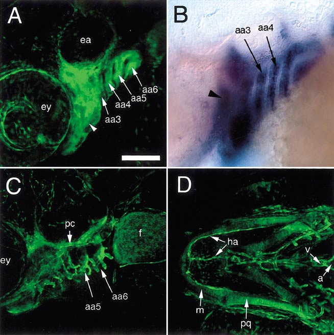
Fig. 3 EGFP expression in cranial neural crest derivatives in TG(fli1:EGFP)y1; albb4 embryos. (A–C) Lateral views, anterior is to the left, dorsal is up. (A) Live 2-dpf embryo expressing EGFP within the aortic arches (aa3–6aa, arrows) and the mesenchyme of the forming jaw (arrowhead); ey, eye; ea, ear. (B) Whole-mount in situ hybridization of endogenous fli1 transcript showing expression within aortic arches (arrows) and jaw primordium (arrowhead, out of focal plane). (C) Live 7-dpf larva showing EGFP expression in structures such as the parachordal cartilage (pc) as well as the vessels within the gills arches (aa5 and aa6; white arrow). EGFP expression is also apparent in the mesenchyme of the developing fin bud (f). (D) Ventral view of embryo in (C); EGFP expression in cells of the forming the jaw, such as Meckel’s cartilage (m) and the palatoquadrate (pq), and associated vessels, such as the hypobranchial artery (ha) as well as the ventricle (v) and atrium (a) of the heart. Scale bar, 100 μm.
Reprinted from Developmental Biology, 248(2), Lawson, N.D. and Weinstein, B.M., In vivo imaging of embryonic vascular development using transgenic zebrafish, 307-318, Copyright (2002) with permission from Elsevier. Full text @ Dev. Biol.