
|
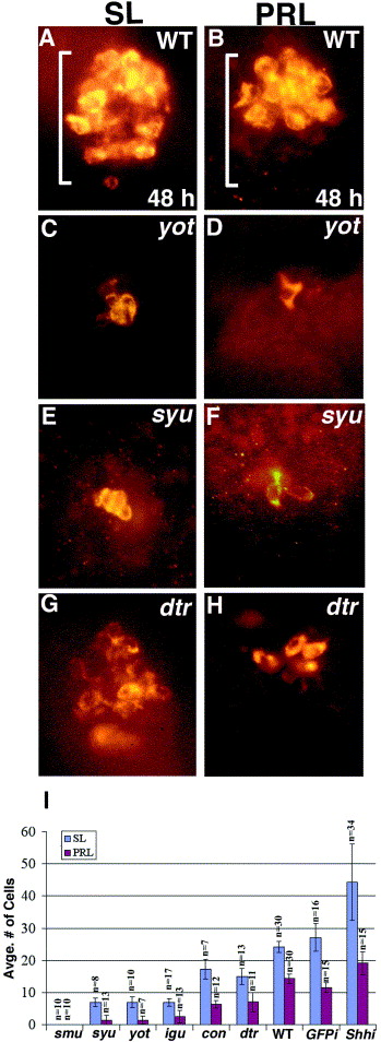
Fig. 7 Reduction of Prolactin (PRL)- and Somatolactin (SL)-secreting cells in Hh pathway mutants. (A, B) In wild-type embryos, SL-secreting cells are present throughout the adenohypophysis, while PRL-secreting cells are only present anteriorly. (C, D) yot/gli2-DR and (E, F) syu/shh mutants have severely reduced numbers of SL- and PRL-secreting cells. (G, H) SL and PRL cells are more moderately reduced in dtr/gli1 mutant embryos. (I) Bar graph showing average numbers of SL and PRL cells in all of the mutants examined ± s.d., numbers of embryos indicated by n. Rightmost bars show numbers of cells present at 30 h following injection of 100 pg of shh mRNA at the two- to four-cell stage. (A–H) Ventral views of 48-h embryos. Brackets show anterior/posterior extent of the adenohypophysis in wildtype embryos.
Reprinted from Developmental Biology, 254(1), Sbrogna, J.L., Barresi, M.J., and Karlstrom, R.O., Multiple roles for Hedgehog signaling in zebrafish pituitary development, 19-35, Copyright (2003) with permission from Elsevier. Full text @ Dev. Biol.